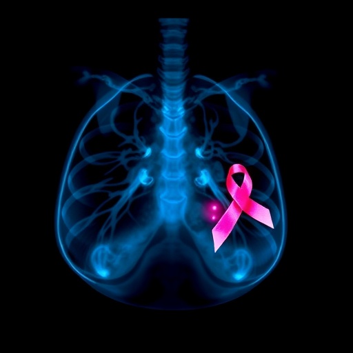
Early detection plays a crucial role in improving survival rates for breast cancer, yet it remains a significant challenge, especially among women with dense breast tissue. Dense breast tissue, prevalent in nearly half of all women in the United States, masks cancer cells during traditional mammographic imaging, making it difficult to spot early tumors. Researchers at Mayo Clinic have conducted a groundbreaking study demonstrating that the integration of molecular breast imaging (MBI) alongside standard 3D mammography can significantly enhance cancer detection rates, particularly in dense breast tissue.
Mammography has long been the cornerstone of breast cancer screening, credited with reducing mortality through early identification of malignancies. However, its effectiveness diminishes in dense breasts owing to the similar radiographic appearances of dense tissue and tumors, which often leads to missed diagnoses. MBI, a functional imaging technique using radiotracers to highlight cancer cells, provides a molecular-level view that circumvents this limitation by distinguishing cancerous tissue based on cellular activity rather than density alone.
In a comprehensive multi-center trial involving nearly 3,000 women aged between 40 and 75 with dense breast tissue, Mayo Clinic researchers compared the efficacy of combined screening using both 3D mammography (digital breast tomosynthesis) and MBI versus each method independently. The dual strategy more than doubled the detection rate of breast cancer within this high-risk group. This enhanced sensitivity was particularly valuable in identifying invasive and aggressive tumors, which might otherwise go unnoticed until reaching advanced stages.
Carrie Hruska, Ph.D., a professor of medical physics and lead author of the study published in the journal Radiology, emphasized the value of early detection for invasive cancers that develop rapidly. “Our research highlights that cancers camouflaged by dense breast tissue can grow undetected with mammography alone. By integrating MBI, we can expose these lethal tumors sooner, potentially saving more lives,” Dr. Hruska explained. This intervention addresses a critical gap in breast cancer screening protocols, marking a milestone in personalized imaging strategies.
The study’s methodology involved annual screenings at five different centers, where each participant received both an MBI scan and a 3D mammogram. The molecular imaging technique utilizes a small dose of a radiotracer that is absorbed by cancer cells, emitting signals captured by the imaging device. Digital breast tomosynthesis, meanwhile, acquires multiple X-ray images from different angles, reconstructing a 3D representation of breast tissue. Together, these complementary imaging modes enhance visualization by combining anatomical detail with functional tumor marker signals.
Notably, the combined approach resulted in increased callback rates in the first year, with 279 additional women requiring further evaluation. Nonetheless, this elevated recall rate was balanced by a significant drop during the second screening cycle, suggesting that the majority of suspicious findings were clarified upon follow-up. This is a critically important observation, as minimizing unnecessary biopsies and anxiety-inducing callbacks maintains the balance between vigilance and overdiagnosis in cancer screening.
Accessibility of MBI combined with mammography has expanded, with about 30 medical sites across the United States offering this advanced diagnostic option. Among these are Mayo Clinic campuses in Rochester, Phoenix, and Jacksonville, as well as Mayo Clinic Health System locations in La Crosse and Eau Claire, Wisconsin. Broader availability means that more women with dense breast tissue can access enhanced screening, improving outcomes through earlier intervention.
Despite its promise, one challenge with MBI lies in the duration of imaging. Current practice requires approximately 40 minutes per scan, which may limit throughput in busy clinical environments and impact patient comfort. Recognizing this, Dr. Hruska’s team is actively working on algorithmic advancements aimed at reducing image acquisition time to around 20 minutes or less. This development could streamline workflow, improve patient experience, and expand eligibility for this supplemental screening modality.
The incorporation of molecular breast imaging alongside traditional mammography represents a paradigm shift in breast cancer detection tailored to individual tissue characteristics. By integrating functional and structural information, clinicians gain a more complete understanding of breast pathology, leading to earlier diagnosis of fast-growing or otherwise occult tumors. Given that breast cancer remains one of the leading causes of cancer-related deaths among women worldwide, innovations like this have the potential to significantly improve survival statistics.
Technical advancements in imaging technology are rapidly evolving, and the concept of combining modalities to overcome the limitations of each is becoming a hallmark of modern diagnostic radiology. Screening programs may increasingly adopt personalized protocols influenced by breast density, genetic risk factors, and imaging responsiveness. The Density MATTERS trial, as the study is named, exemplifies this tailored approach by addressing a key variable affecting mammographic sensitivity.
Ultimately, patient education about breast density and supplemental screening options is paramount. While mammography remains indispensable as a baseline screening tool, awareness of additional tests like MBI can empower women to seek comprehensive evaluation based on their individual risk profile. Healthcare providers, radiologists, and oncologists must work collaboratively to effectively communicate these advances and their implications for screening frequency and modality selection.
In summary, the combination of molecular breast imaging with digital breast tomosynthesis significantly improves early detection of breast cancer in women with dense breast tissue by enhancing visualization of invasive tumors. This dual-imaging strategy offers a promising avenue to overcome the limitations of traditional mammography, reduce advanced-stage diagnosis, and improve survival outcomes. As technological refinements continue and accessibility expands, this approach may become a new standard of care for breast cancer screening in dense breasts.
Subject of Research: Breast cancer detection improvements in women with dense breast tissue using combined molecular breast imaging and 3D mammography
Article Title: Molecular Breast Imaging and Digital Breast Tomosynthesis for Dense Breast Screening: The Density MATTERS Trial
News Publication Date: 23-Sep-2025
Web References:
– https://www.mayoclinic.org/diseases-conditions/breast-cancer/symptoms-causes/syc-20352470
– https://newsnetwork.mayoclinic.org/discussion/mayo-clinic-q-and-a-breast-density-reporting-and-supplemental-testing/
– https://newsnetwork.mayoclinic.org/discussion/mayo-clinic-minute-molecular-breast-imaging-for-supplemental-breast-screening/
– https://www.mayoclinic.org/tests-procedures/3d-mammogram/about/pac-20438708
References:
– Hruska, C., et al. Molecular Breast Imaging and Digital Breast Tomosynthesis for Dense Breast Screening: The Density MATTERS Trial. Radiology. (Published 23-Sep-2025). https://pubs.rsna.org/doi/10.1148/radiol.243953
Keywords:
Breast cancer, dense breast tissue, molecular breast imaging (MBI), digital breast tomosynthesis, 3D mammography, breast cancer screening, early detection, radiology, diagnostic imaging, Mayo Clinic, breast cancer detection, supplemental screening
