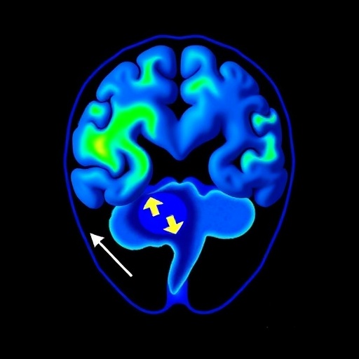
In a groundbreaking advancement at the crossroads of neuroscience and artificial intelligence, researchers have unveiled an innovative approach to understanding the aftermath of hypothalamic hamartoma (HH) surgery through multimodal contrastive learning applied to resting-state functional MRI (rs-fMRI) data. This new technique reveals subtle yet significant changes in the brain’s functional networks, offering promising insights into whole-brain network recovery—a feat that traditional neuroimaging analyses have long struggled to achieve.
Hypothalamic hamartomas, congenital malformations located near the hypothalamus, are notorious for inducing severe epilepsy that frequently resists pharmacological treatment. Surgical removal of HH is often the only viable option to control seizures but assessing how this intervention affects brain-wide network function has posed a formidable challenge. Conventional rs-fMRI analyses encounter limitations in detecting minute but critical shifts in the complex interplay of neural circuits post-surgery, obscuring a full picture of cerebral recovery.
Addressing this challenge head-on, a team led by Jeyabose and colleagues developed a sophisticated two-stage contrastive learning algorithm capable of discerning intricate network changes by integrating multi-dimensional rs-fMRI data. This approach uniquely combines spatial and temporal information—specifically three-dimensional Independent Component Analysis (ICA) maps with one-dimensional ICA time series—allowing the model to encode rich, multifaceted representations of brain activity before and after surgery.
The first stage of their model functions as a multimodal contrastive encoder, differentiating pre-operative and post-operative states across disparate functional domains such as motor, vision, language, frontal, and temporal networks. By leveraging contrastive objectives, the encoder simultaneously learns to maximize distinctions between these states while preserving meaningful network-specific characteristics. This ensures that embeddings not only separate conditions but also maintain fidelity to the underlying neural substrates.
Subsequently, a lightweight classifier refines these learned embeddings, augmented by the original ICA inputs, to deliver precise network-wise classifications. This hierarchical methodology enhances sensitivity and specificity in capturing subtle functional transitions, surpassing the limitations of traditional statistical analyses often prone to averaging out critical neural dynamics or missing nuanced patterns altogether.
Visual inspection of the learned feature space via t-distributed stochastic neighbor embedding (t-SNE) revealed stark separation between pre-surgical and post-surgical brain states. This clear delineation across all five examined networks underscores the model’s capacity to identify functional reorganization induced by surgical intervention—a milestone in neuroengineering that bridges computational sophistication with clinical applicability.
Quantitative evaluation of the model displayed impressive performance metrics: classification accuracy ranged from 85% to 90%, sensitivity spanned 79% to 90%, and specificity ranged between 87% and 93%. The F1-scores and area under the curve (AUC) values similarly indicated robust discriminative power, affirming the reliability and consistency of these neural biomarkers in reflecting postoperative recovery.
These findings herald a new era where advanced machine learning frameworks can sensitively detect cerebral adaptations post-HH surgery, providing unprecedented biomarkers for epileptic encephalopathy and recovery tracking. By illuminating changes in motor, vision, language, frontal, and temporal cortical networks, the research paves the way for real-time, non-invasive monitoring strategies that clinicians can employ to personalize treatment trajectories and optimize patient outcomes.
Beyond immediate clinical implications, this study exemplifies how multimodal neuroimaging data, when paired with cutting-edge contrastive learning paradigms, can unravel the intricate dynamics of brain connectivity with unmatched resolution. Such methodologies may revolutionize the study of brain plasticity, neurorehabilitation, and the broader spectrum of neurological disorders where network dysfunction plays a pivotal role.
Moreover, the authors advocate for future work to extend these analytic frameworks by including healthy control cohorts. This would enable comparative studies to quantify objective markers of network recovery and resilience, deepening our understanding of how pathological brain states normalize or reorganize following interventions. These comparative analyses could provide foundational knowledge for developing novel prognostic tools and therapeutic targets.
On a technical front, the blend of spatial and temporal ICA data feeding into the contrastive learning architecture represents an elegant marriage of data modalities. This integrative paradigm ensures that both the static and dynamic dimensions of brain function are captured, reflecting the complex, time-evolving nature of neural circuitry. Such comprehensive encoding strategies are critical for advancing neuroimaging analytics beyond conventional snapshots of brain activity.
Collectively, this pioneering research signifies a paradigm shift in epilepsy surgery evaluation, where artificial intelligence transcends mere pattern recognition to offer mechanistic insights into brain recovery. The implications resonate across neuroengineering, clinical neuroscience, and computational neurology, inspiring a future where precise network-tailored treatments become a tangible reality.
As researchers continue refining these algorithms, integrating multimodal datasets promises to unlock deeper mysteries of brain function and plasticity. With every step, the convergence of machine learning and neuroscience edges closer to delivering transformative clinical innovations that can restore lives disrupted by intractable neurological conditions like hypothalamic hamartoma-associated epilepsy.
Subject of Research:
The study focuses on quantifying whole-brain network recovery after hypothalamic hamartoma surgery using multimodal contrastive learning applied to resting-state functional MRI data.
Article Title:
Multimodal contrastive learning on rs-fMRI to quantify whole-brain network recovery after hypothalamic hamartoma surgery.
Article References:
Jeyabose, A., Robinson, B., Boerwinkle, V.L. et al. Multimodal contrastive learning on rs-fMRI to quantify whole-brain network recovery after hypothalamic hamartoma surgery. BioMed Eng OnLine 24, 125 (2025). https://doi.org/10.1186/s12938-025-01458-6
Image Credits: AI Generated
DOI:
https://doi.org/10.1186/s12938-025-01458-6


