


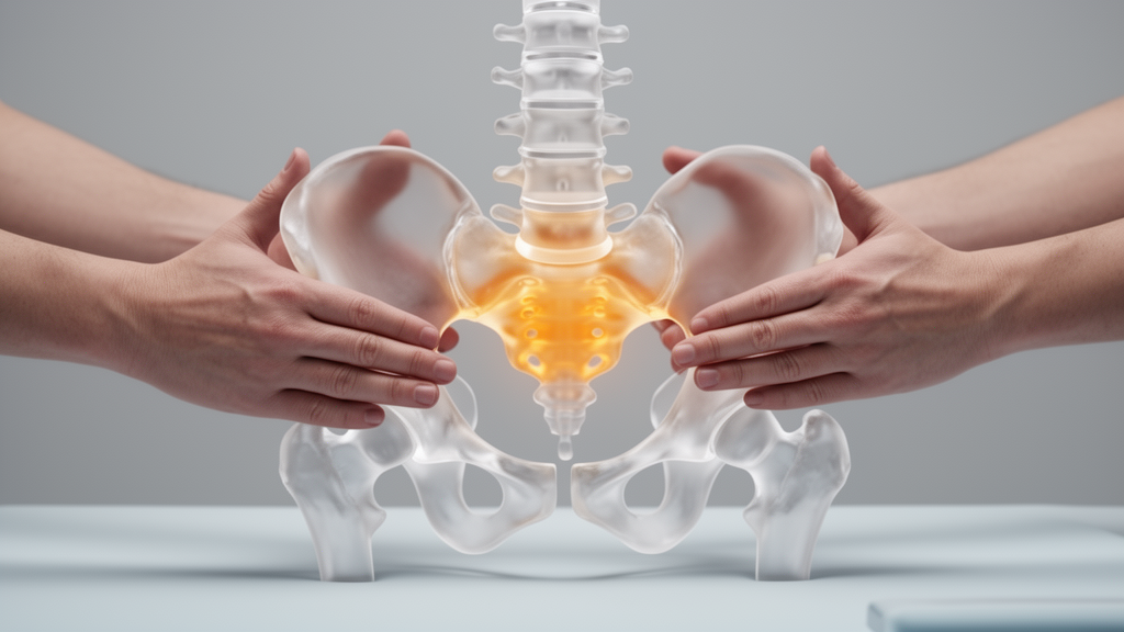
For millions of people living with stubborn low back pain, the sacroiliac joint has long been a shadowy suspect. Tucked deep between the sacrum and the pelvis, this joint is increasingly recognized as a pain generator in roughly 15 to 30 percent of low back pain cases, yet its clinical presentation overlaps so heavily with disc and facet joint syndromes that it is frequently overlooked. Because sacroiliac joint dysfunction is usually managed without surgery, manual therapy remains a mainstay of conservative care. But a fundamental question has divided clinicians and biomechanists for decades: when a therapist performs a manipulation, is the benefit coming from actually mobilizing the joint itself, or from modulating the soft tissues wrapped around it? A new computational study from a team at Guangdong Provincial Hospital of Chinese Medicine and the Chinese Medicine Guangdong Laboratory offers some of the most detailed answers yet, and the findings are striking.
The research, published in Frontiers in Bioengineering and Biotechnology, used three-dimensional finite element analysis to compare how the sacroiliac joint and the iliolumbar ligament respond to four distinct loading configurations: physiological compressive stress, traction manipulation stress, oblique-pulling manipulation, and counter-directional sacral thrust with iliac mobilization. Finite element analysis is a computational technique that breaks a complex structure into millions of tiny elements, allowing engineers and biomechanists to calculate stress, deformation, and motion at locations no physical sensor could reach. In this case, the team reconstructed a lumbar spine-pelvis model from computed tomography data of a healthy 31-year-old male volunteer, generating a model of extraordinary resolution: more than two million nodes and nearly 1.27 million elements.
The technical rigor of the model deserves attention. Cortical bone was assigned an elastic modulus of 12,000 megapascals, while cancellous bone received just 100 megapascals, reflecting the dramatic difference between dense outer shell and porous inner structure. The sacroiliac joint cartilage, approximately two millimeters thick, was modeled as a solid with an elastic modulus of 100 megapascals. The iliolumbar ligament was reconstructed as six irregular three-dimensional solid bundles following anatomical landmarks, extending from the lower lumbar transverse processes to their attachments along the iliac crest. Other sacroiliac ligaments, including the anterior sacroiliac, posterior sacroiliac, interosseous sacroiliac, sacrotuberous, and sacrospinous ligaments, were represented using tension-only spring elements with stiffness values ranging from 400 to 2,800 newtons per millimeter. The interosseous sacroiliac ligament, at 2,800 newtons per millimeter, was the stiffest of them all, underscoring how tightly constrained this joint really is.
Before running the four loading scenarios, the team validated their model against previously published cadaveric and finite element data. They simulated a single-leg stance condition by fixing the left ilium and applying a 7.5 newton-meter moment to the superior endplate of L4, reproducing flexion-extension, lateral bending, and axial rotation. The calculated sacroiliac joint range of motion values were generally within or close to the reported ranges from Lindsey and colleagues and Zhang and colleagues, with only mild deviations in left sacroiliac joint flexion-extension and axial rotation. A three-level local cartilage mesh assessment under the oblique-pulling condition showed that refining the mesh from 1.0 to 0.5 millimeters changed peak cartilage stress by only 2.68 percent, indicating that further refinement would yield diminishing returns. The stress maxima remained in the same anatomical region across all three mesh densities.
The four loading conditions were then simulated with careful attention to the prescribed terminal states. Physiological compressive stress involved fully constraining both iliac sides and applying a 300 newton downward force to the superior endplate of L4 along the direction of body weight. Traction manipulation stress reversed that force, applying 300 newtons upward to simulate the mechanical effect of traction on the joint. Oblique-pulling manipulation combined 300 newtons of upward traction at L4 with a prescribed 10-degree posterior rotation of the right ilium through a remote displacement boundary condition, a terminal angle selected based on previous cadaveric studies. Counter-directional sacral thrust with iliac mobilization applied a 600 newton posterior force to the surface of the right ilium along the sacroiliac joint direction, a load selected based on previous measurements of manipulation forces recorded from five therapists averaging approximately 600 newtons.
The results revealed a hierarchy of mechanical responses that could reshape how clinicians think about these techniques. Physiological compressive stress and traction manipulation stress produced nearly identical cartilage responses, with peak von Mises stress values of 1.456 and 1.457 megapascals respectively, and a peak total deformation of just 0.041 millimeters under both conditions. Simply reversing a 300 newton axial load under bilateral iliac constraint produced almost no change. In stark contrast, oblique-pulling manipulation generated the highest peak cartilage stress and deformation at 35.174 megapascals and 0.553 millimeters, followed by counter-directional sacral thrust with iliac mobilization at 11.057 megapascals and 0.209 millimeters. The range of motion of the right sacroiliac joint reached 0.62 degrees under oblique-pulling manipulation and 0.21 degrees under counter-directional sacral thrust, compared with just 0.02 degrees under the baseline conditions.
Principal-stress analysis added another layer of insight. The maximum principal stresses in the cartilage were 1.5476, 1.6921, 21.256, and 5.938 megapascals under the four conditions, while the corresponding minimum principal stresses were −1.7196, −1.5554, −47.767, and −15.252 megapascals. Oblique-pulling manipulation therefore produced the largest positive and negative cartilage stress extrema, indicating both strong tensile and strong compressive components within the joint. The peak maximum principal stress under oblique-pulling manipulation was located at the lateral cartilage margin, where the local deformation pattern was consistent with lateral bulging under compression. Perhaps most intriguingly, the surface showing greater cartilage deformation changed depending on the loading configuration. Under physiological and traction loading, the sacral cartilage surface showed greater deformation than the iliac surface on the right side. Under both manipulative conditions, this relationship reversed, with the iliac surface showing greater deformation. Under oblique-pulling manipulation, right iliac-side deformation was more than twice the right sacral-side value.
The iliolumbar ligament told its own story. Peak ligament stress values were 4.106, 4.108, 4.717, and 1.306 megapascals under the four conditions, while corresponding deformation values were 1.529, 1.530, 1.604, and 0.451 millimeters. Oblique-pulling manipulation generated the greatest ligament stress and deformation, with the response most evident in the right upper bundle. Counter-directional sacral thrust with iliac mobilization, by contrast, produced the lowest ligament response among all four conditions, reducing peak ligament stress and deformation by 68.19 percent and 70.52 percent relative to physiological loading. Maximum directional displacement along the assigned local X-axis was greatest under oblique-pulling manipulation for all six bundles, with the right upper, right middle, and right lower bundles reaching 0.71854, 0.59354, and 0.58541 millimeters respectively. These direction-specific results indicate that oblique-pulling manipulation produced the most pronounced ligament motion along the principal loading direction, particularly in the right-sided bundles.
What does this mean for the long-standing controversy? The findings suggest that oblique-pulling manipulation and counter-directional sacral thrust with iliac mobilization represent mechanically distinct maneuvers rather than a single mechanism operating at different intensities. Oblique-pulling manipulation combines upward loading at L4 with posterior rotation of the right ilium, creating a coupled rotation-traction pattern associated with a greater iliolumbar ligament response. Counter-directional sacral thrust applies a posterior force along the joint direction and appears to modify the articular response with relatively limited ligament response. The absolute joint motion remained within a sub-degree range even under the manipulative conditions, compatible with the small angular magnitudes reported for intact sacroiliac joints under physiological and experimental loading. This aligns with previous in vivo evidence, including a roentgen stereophotogrammetric analysis by Tullberg and colleagues that found no measurable positional change in the sacroiliac joint after manipulation.
However, limited joint motion does not imply limited mechanical relevance. The sacroiliac joint operates as a constrained load transfer joint whose stability depends on joint congruence, articular surface morphology, frictional resistance, and strong ligamentous constraints, a mechanism described as self-bracing and later developed into the framework of form closure and force closure. Even sub-degree motion may affect joint constraint and ligament response, with potential implications for lumbopelvic load transmission. Cadaveric research has demonstrated that sequential transection of the iliolumbar ligament significantly increases sagittal sacroiliac joint mobility, indicating that ligament restraint contributes directly to joint constraint. Immunohistochemical studies have identified nociceptive structures in sacroiliac joint cartilage and adjacent ligamentous tissues, suggesting that local stress redistribution may be clinically relevant even when observable joint motion remains small.
The clinical literature broadly supports the idea that manual therapy can improve outcomes in sacroiliac joint pain syndrome without requiring radiographically demonstrable joint repositioning. A recent systematic review and meta-analysis concluded that manual therapy may improve disability in sacroiliac joint pain syndrome while emphasizing heterogeneity in the current evidence base. Randomized controlled trials have reported that both manipulation and exercise therapy improved outcomes in patients with sacroiliac joint dysfunction, and other studies found that manual therapy combined with sacroiliac and lumbar exercises improved pain and function in selected patients. None of these studies demonstrated that clinical success required verifiable joint resetting, a finding compatible with the limited joint motion and altered periarticular mechanical responses observed in the present model.
The study has limitations that the authors acknowledge candidly. The model was based on one healthy adult male and cannot represent age-, sex-, or pathology-related variability. The clinical maneuvers were represented by simplified terminal loading conditions rather than their full dynamic processes. The static model omitted active muscle forces and modeled sacroiliac joint cartilage as linear elastic without depth-dependent, anisotropic, biphasic, or time-dependent behavior. The iliolumbar ligaments were simplified as linear isotropic solids without pretension or time-dependent behavior, and ligament pretension may influence model outputs. The frictionless, no-separation contact definition and rigid sacral fixation represent additional modeling assumptions. Future analyses incorporating alternative contact formulations, compliant support conditions, higher-resolution imaging, and additional validation data may further clarify the anatomical and mechanical representation of subject-specific sacroiliac joint structures.
Nevertheless, the findings represent a significant step toward understanding what actually happens inside the sacroiliac joint during manual therapy. By showing that oblique-pulling manipulation produces a more pronounced concurrent articular-ligamentous response while counter-directional sacral thrust generates increased articular responses with comparatively smaller iliolumbar ligament responses, the study provides a biomechanical framework for distinguishing between techniques that have long been treated as interchangeable. The reversal in which cartilage surface shows greater deformation, from sacral to iliac, offers a surface-specific signature that could guide future experimental validation and technique refinement. For a joint that has resisted direct measurement for decades, computational models like this one may be the clearest window yet into the mechanical world of manual therapy.
Subject of Research: Effects of physiological and manipulative loading conditions on the sacroiliac joint: a three-dimensional finite element analysis
Article Title: Effects of physiological and manipulative loading conditions on the sacroiliac joint: a three-dimensional finite element analysis
Article References: Ma, J., Zhou, X., Xu, S., Dai, L., Wang, Y., Lin, J., Lin, D., & Qi, J. (2026). Effects of physiological and manipulative loading conditions on the sacroiliac joint: a three-dimensional finite element analysis. Frontiers in Bioengineering and Biotechnology, 14, Article 1906528. https://doi.org/10.3389/fbioe.2026.1906528
Image Credits: AI Generated
DOI: 10.3389/fbioe.2026.1906528
Keywords: Effects, physiological, manipulative, loading, conditions, sacroiliac, joint, three-dimensional, finite, element, analysis, scientific research
Cite Scienmag News
Denise Maddox. (October 19, 2026). Manipulation or soft tissue? Finite element model reveals how manual therapy loads the sacroiliac joint. Scienmag. https://scienmag.com/manipulation-or-soft-tissue-finite-element-model-reveals-how-manual-therapy-loads-the-sacroiliac-joint/
Denise Maddox. "Manipulation or soft tissue? Finite element model reveals how manual therapy loads the sacroiliac joint." Scienmag, 19 October 2026, https://scienmag.com/manipulation-or-soft-tissue-finite-element-model-reveals-how-manual-therapy-loads-the-sacroiliac-joint/. Accessed 19 October 2026.
Denise Maddox. "Manipulation or soft tissue? Finite element model reveals how manual therapy loads the sacroiliac joint." Scienmag. October 19, 2026. https://scienmag.com/manipulation-or-soft-tissue-finite-element-model-reveals-how-manual-therapy-loads-the-sacroiliac-joint/


