


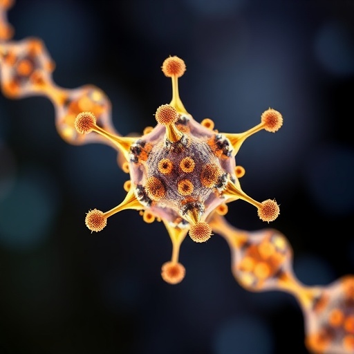
Nanoplastics may be almost impossible to see, but that does not make them biologically insignificant. Particles smaller than a micrometre can move through environmental systems, enter organisms and cross biological barriers, yet scientists have struggled to determine exactly where they go, how much accumulates and how long it remains there. A new study describes a machine-learning-designed nanoplatform that could make these movements far easier to measure. The technology combines fluorescence imaging with an independent metal-based signal, allowing researchers to track nanoplastics in space and time while correcting for one of fluorescence microscopy’s most serious weaknesses: signal decay. In experiments reported in Nature Sensors, the bifunctional nanoprobes achieved more than 90 per cent recovery in complex matrices and revealed greater cellular uptake, longer retention and more extensive transfer between generations than fluorescence-only measurements detected. The results suggest that conventional methods may be systematically underestimating the persistence and biological reach of nanoplastics.
The central problem is a mismatch between visual detail and chemical accuracy. Fluorescent labels can show where a particle is located inside a cell or organism, often with excellent spatial resolution. But fluorescent molecules can fade under illumination, a phenomenon known as photobleaching. Their signal can also be altered by pH, polarity, local molecular crowding or interactions with biological material. As a result, a dimmer signal does not necessarily mean that fewer particles remain. It may simply mean that the label has degraded or that its optical environment has changed. Conversely, measuring the total amount of plastic in a sample can provide stronger quantitative information but generally loses the fine-scale information needed to identify which cells or tissues contain the particles. This trade-off becomes especially damaging in long-term studies, where researchers need to compare locations and concentrations across hours, days or generations. The new strategy was designed to preserve the detailed information supplied by imaging while adding a more stable internal reference for quantification.
The researchers used machine learning to guide the design of nanoprobes with two complementary reporting functions. One component produces fluorescence through aggregation-induced emission, a process in which molecules become strongly emissive when constrained in close-packed assemblies. Unlike many conventional fluorescent dyes, aggregation-induced-emission materials can be designed to remain bright when clustered, making them useful for constructing nanoscale tracking labels. The second component incorporates stable metal dopants into the same probe. These metals do not provide a conventional optical image; instead, they act as a chemically measurable signal whose abundance is not expected to fade in the same way as fluorescence. The resulting particle therefore carries both a visual beacon and a quantitative anchor. Machine-learning-assisted design was used to identify probe compositions and structures that could balance these functions, improving the chance that the fluorescent and metal signals would remain correlated as the particles moved through complicated biological and environmental settings.
This dual-signal architecture creates what the researchers describe as a self-calibrated measurement system. Fluorescence reports where the nanoprobes are and can provide high-resolution maps of their distribution. The metal signal supplies a time-invariant reference that can be measured independently. If the fluorescent intensity falls during prolonged observation, the metal measurement can help determine whether the decline reflects genuine loss of particles or merely optical fading. In practical terms, the metal channel functions like an internal ruler for the fluorescence channel. By combining the two signals with the time of observation, researchers can build a four-dimensional framework linking fluorescence, metal content, time and concentration. That framework is important because nanoplastic exposure is not a static event. Particles can be taken up, transported, stored, released or passed onward, and each process changes their concentration in particular tissues or cells. A method that records only one moment or one signal can miss these transitions.
Testing in complex matrices is crucial because nanoplastics behave differently in purified water, cell cultures and living organisms. Proteins, salts, lipids and other organic molecules can coat particle surfaces, alter their aggregation and interfere with detection. Biological tissues can also absorb or scatter light, complicating fluorescence measurements and making it difficult to recover every particle introduced into a sample. According to the study, the bifunctional nanoprobes achieved recovery rates above 90 per cent in such complex conditions. That result indicates that the probes’ metal-based quantification remained effective even when the surrounding material could distort optical signals. High recovery does not by itself establish how every environmental nanoplastic behaves, because the engineered probes are measurement tools rather than a complete catalogue of naturally occurring particles. It does, however, demonstrate that the platform can retain quantitative performance where fluorescence alone is most vulnerable. That distinction could prove decisive for experiments designed to follow exposure over extended periods.
When the researchers compared the dual-signal approach with fluorescence-only tracking, the difference was biologically consequential. The internally calibrated measurements indicated that cells took up more nanoplastic material than fluorescence measurements alone suggested. They also showed that particles remained inside biological systems for longer periods and that transfer between generations was more extensive than previously detected through the optical signal alone. These observations are consistent with a familiar problem in microscopy: once a fluorescent label fades, the object may appear to have disappeared even though its nonfluorescent components are still present. In a short experiment, that loss may be a technical inconvenience. In a long-term exposure study, it can change the apparent biological conclusion, turning persistent particles into apparently transient ones. By retaining the metal signal as a stable reference, the new platform can distinguish a real reduction in particle abundance from a reduction in detectability. That distinction strengthens estimates of accumulation and retention, two factors that influence the potential risk of chronic exposure.
The possibility of transgenerational transfer makes the measurement challenge even more important. If nanoplastic material remains in an organism long enough to enter reproductive pathways or developing offspring, exposure may continue after the original environmental contact has ended. Detecting such transfer requires more than showing that fluorescent particles are visible in a parent and later in descendants. Researchers must establish how much material is present, where it is located and whether a changing signal reflects movement or label degradation. The study’s four-dimensional framework is intended to address those questions by linking spatial observations to calibrated concentration and elapsed time. The reported evidence of greater long-term retention and transgenerational transfer than fluorescence alone revealed does not mean that every nanoplastic will behave identically, nor does it by itself define a health effect. It does show that the apparent duration and scale of exposure can depend strongly on the analytical method used to measure it.
The platform may also help connect laboratory observations with broader environmental risk assessment. Nanoplastics arise as larger plastic materials weather and fragment, and their small size gives them access to biological interfaces that larger particles cannot cross as easily. Yet risk estimates require reliable information about dose, residence time and tissue distribution—parameters that are difficult to obtain when the signal used for tracking is unstable. A probe that combines microscopy with an elemental or metal-based measurement could be adapted to study interactions between nanoscale materials and cells, tissues or whole organisms. The approach may also be relevant beyond plastics, wherever researchers need to follow particles over long periods in optically complex environments. Its value will depend on further validation, including how closely the engineered nanoprobes reproduce the surface chemistry, shape and behavior of real environmental nanoplastics. The study establishes a measurement strategy, not a final answer to the health and ecological questions surrounding plastic pollution.
The most important implication is methodological: what scientists cannot reliably measure may be mistaken for what does not persist. By correcting fluorescence with a stable internal signal, the new nanoprobes offer a way to observe nanoscale contaminants without accepting the usual compromise between spatial resolution and quantitative accuracy. The machine-learning-guided design further suggests that optical and chemical measurements can be engineered together rather than treated as separate assays. If the system performs consistently across different organisms, tissues and environmental conditions, it could sharpen studies of how nanoplastics cross barriers, accumulate in cells and move through generations. For now, the findings provide a warning against interpreting a fading fluorescent image as proof that a particle has vanished. Nanoplastics may be more persistent, more widely distributed and more biologically mobile than standard imaging has shown—and a more dependable measuring stick could be the first step toward discovering just how extensive their hidden journey is.
Cite Scienmag News
Denise Maddox. (August 27, 2026). Machine-learning-designed bifunctional nanoprobes enable self-calibrated tracking of nanoplastics across space and time. Scienmag. https://scienmag.com/machine-learning-designed-bifunctional-nanoprobes-enable-self-calibrated-tracking-of-nanoplastics-across-space-and-time/
Denise Maddox. "Machine-learning-designed bifunctional nanoprobes enable self-calibrated tracking of nanoplastics across space and time." Scienmag, 27 August 2026, https://scienmag.com/machine-learning-designed-bifunctional-nanoprobes-enable-self-calibrated-tracking-of-nanoplastics-across-space-and-time/. Accessed 6 September 2026.
Denise Maddox. "Machine-learning-designed bifunctional nanoprobes enable self-calibrated tracking of nanoplastics across space and time." Scienmag. August 27, 2026. https://scienmag.com/machine-learning-designed-bifunctional-nanoprobes-enable-self-calibrated-tracking-of-nanoplastics-across-space-and-time/


