


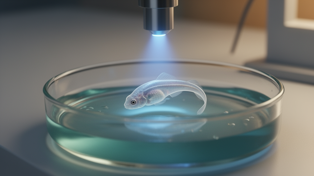
Neuroscientists have long dreamed of controlling the brain the way a conductor controls an orchestra: precisely, reversibly, and without permanently rewiring the instrument. A new study brings that dream a step closer, showing that a single light-activated molecule can disrupt a well-studied sensorimotor behavior in zebrafish larvae — and then hand control back the moment the light changes. The work, published in Frontiers in Behavioral Neuroscience, marks the first time that the zebrafish optomotor response, one of the field’s most trusted behavioral assays, has been combined with photopharmacology in wildtype animals.
The compound at the center of the study is Carbadiazocine, a photoswitchable analog of carbamazepine, a widely used anticonvulsant that blocks voltage-gated sodium channels and thereby controls neuronal firing. What makes Carbadiazocine special is that it exists in two interconvertible forms, or photoisomers. In its dark-relaxed state, the molecule is essentially inert: adding it to the water of zebrafish larvae has no effect on their locomotion. But when illuminated with 405-nanometer violet light, roughly half the molecules flip into an active configuration that changes their biological effect. Irradiation with a second wavelength, 500 nanometers, can push them back. This isomerization is reversible with time as the compound thermally relaxes, or immediately upon irradiation with the second wavelength, giving researchers a molecular dial they can turn on and off with light.
Photopharmacology, the discipline behind such compounds, is often described as optogenetics’ quieter sibling. Where optogenetics requires genetically modifying animals to express light-sensitive proteins such as channelrhodopsin, photoswitchable drugs work on endogenous proteins in their physiological context. Any wildtype organism expressing the target protein can be controlled, without genetic manipulation. And because the drug’s activity can be constrained spatially by illumination patterns, researchers can in principle activate it only in the brain region of interest, minimizing off-target effects and adverse consequences elsewhere. The approach has already been shown to modulate larval zebrafish locomotion and to alleviate neuropathic pain in rodents in a reversible, light-induced manner.
To test whether this molecular dial could reach the circuits that translate vision into movement, the team — led by Luisa Camerin and Joaquín Martínez-Tambella of the Institute for Bioengineering of Catalonia and the University of Barcelona, together with Gregor Schuhknecht, Vickie M. Wang, Kumaresh Krishnan, and Paula Pflitsch working in Florian Engert’s laboratory at Harvard, under the supervision of Engert and Pau Gorostiza — turned to the optomotor response, or OMR. Zebrafish possess an innate ability to detect visual motion and swim in the direction of perceived movement, a survival mechanism thought to help them avoid involuntary displacement in water currents and evade predators. The behavior requires no training and is present in larvae as early as five days post-fertilization, making it an ideal system for exploring the neural basis of sensorimotor integration and decision-making.
The researchers ran their experiments in two complementary configurations. In the free-swimming paradigm, larvae swam in multiwell plates above a screen projecting high-contrast parallel stripes that moved up and down, and a commercial tracking system recorded their positions. In the head-fixed paradigm, larvae were embedded in transparent agarose with their tails free to move, while a random dot kinematogram — a visual stimulus originally developed for primate studies — projected roughly 1,000 red dots moving perpendicular to the fish body axis in a closed loop with the animals’ movements. Varying the percentage of dots moving coherently, from 0 to 25 to 100 percent, allowed the team to probe how strongly the larvae integrated sensory evidence before committing to a directional decision.
The results were striking. In free-swimming larvae, treatment with pre-illuminated Carbadiazocine caused a two-fold increase in swim distance and a three-fold decrease in correctness — the percentage of larvae moving in the same direction as the visual stimulus dropped from roughly 65 percent to about 20 percent. In head-fixed larvae, correctness fell from about 95 percent to about 80 percent at the strongest stimulus coherence. Crucially, the dark-relaxed isomer produced none of these effects, confirming that the behavioral changes were entirely light-dependent. The hyperactivity was evident immediately upon administration, even before visual stimulus onset, and treated larvae continued to modify their swimming direction each time the stimulus changed — albeit sometimes in the opposite direction to the moving stripes.
Speed analysis revealed that the compound did more than simply increase locomotion. Pre-illuminated Carbadiazocine increased the total number of movement events across all speed categories, but it shortened and reduced the distance covered by slow movements — those below 2 millimeters per second — while making fast movements, above 6 millimeters per second, more frequent and prolonged. In effect, the drug shifted larval displacement from predominantly slow exploratory swimming to a behavior dominated by fast bursts, even in the absence of visual stimuli to guide behavior. Tail bout analysis in head-fixed larvae added a further layer: the compound increased the proportion of swims with tail movement frequencies between 15 and 30 hertz, corresponding to irregular tail movements, and induced a rise in subthreshold small swims below the detection threshold of the tracking software, interspersed with periods of complete inactivity that may reflect exhaustion following hyperactivity.
Interpreting these findings requires appreciating the distributed neural architecture of the optomotor response. Visual motion is first detected by the retina and relayed via retinal arborization fields to the pretectum and optic tectum, where directional information is processed and integrated. Descending motor commands then travel through premotor nuclei, including the nucleus of the medial longitudinal fasciculus and reticulospinal neurons, with potential cerebellar modulation, to spinal cord circuits that drive the tail musculature. This circuit also supports the temporal accumulation of sensory evidence over several seconds. The relatively preserved correctness in the head-fixed condition suggests that initial perception and the directional decision based on sensory input remain largely intact after treatment. In contrast, when behavior is assessed over longer timescales in freely swimming larvae, the drug-induced hyperactivity appears to impair sustained performance — as if the animals skidded or overshot — with trajectories that become irregular, orthogonal to, or even opposite the direction of optic flow.
The team also attempted real-time photoswitching experiments, in which the drug was switched between its states while the larvae were swimming. This is technically challenging because changing light itself produces well-documented startle responses that cannot be avoided by blinding procedures, since the optomotor response requires visual perception. Despite this limitation, the researchers observed a significant decrease in OMR correctness after irradiating the plate with 400-nanometer light in larvae treated with dark-relaxed Carbadiazocine, and these differences were reversed by 500-nanometer illumination — proof that the molecular dial can be turned back and forth in a living animal.
The implications extend well beyond zebrafish. Because the drug works on endogenous sodium channels in any wildtype organism, the same approach could in principle probe neuronal circuits underlying behavior in diverse species, using patterns of photostimulation at wide spatiotemporal scales. The spatial resolution of photostimulation is diffraction-limited to a fraction of a micrometer for the ultraviolet and visible wavelengths used in photopharmacology, and previous work has achieved axial resolution of about 5 micrometers with a diffusible ligand and about 1 micrometer on the image plane with a tethered one, allowing control of individual synapses in vivo. In the larval zebrafish brain, a cluster of roughly 300 descending projection neurons connects to the spinal cord, and systematically testing their activity upon Carbadiazocine administration combined with precise light activation would provide deeper insights into which neuronal subpopulations contribute to the different movement types the drug modulates. Unlike optogenetic manipulations, which operate at the cellular scale through available gene promoters, photopharmacology directly controls endogenous proteins in their physiological context, leveraging their signaling pathways and downstream biological processes, including behavioral outcomes. The researchers suggest that head-fixed experiments with focused laser scanning illumination may offer greater versatility to selectively photoswitch the circuits under study while minimizing confounding signals via the retina.
What emerges from this study is a platform: two advanced behavioral assays, freely swimming and head-fixed, now married to a light-controllable drug whose inactive state leaves behavior untouched. The in-depth characterization of Carbadiazocine’s effects — the type of movements affected, their number, duration, distance, and the bout frequency range altered — posits it as a unique tool to study the neural circuits underlying specific movements using patterns of photostimulation. By selectively perturbing and probing neural computations with spatiotemporal precision, researchers hope to crack the intricate networks underlying behavior, one wavelength at a time.
Subject of Research: Photopharmacology
Article Title: Remote control of wildtype zebrafish optomotor response with a photoswitchable drug
Article References: Camerin, L., Martínez-Tambella, J., Schuhknecht, G., Wang, V. M., Krishnan, K., Pflitsch, P., Engert, F., & Gorostiza, P. (2026). Remote control of wildtype zebrafish optomotor response with a photoswitchable drug. Frontiers in Behavioral Neuroscience, 20, Article 1916443. https://doi.org/10.3389/fnbeh.2026.1916443
Image Credits: AI Generated
DOI: 10.3389/fnbeh.2026.1916443
Keywords: photopharmacology, zebrafish, optomotor response, neuroscience, photoswitchable drug, Carbadiazocine, sensorimotor behavior, circuit perturbation, Remote, control, wildtype, optomotor
Cite Scienmag News
Cassandra Pierce. (October 17, 2026). Light-Activated Drug Steers Zebrafish Behavior Without Touching Their Genes. Scienmag. https://scienmag.com/light-activated-drug-steers-zebrafish-behavior-without-touching-their-genes/
Cassandra Pierce. "Light-Activated Drug Steers Zebrafish Behavior Without Touching Their Genes." Scienmag, 17 October 2026, https://scienmag.com/light-activated-drug-steers-zebrafish-behavior-without-touching-their-genes/. Accessed 17 October 2026.
Cassandra Pierce. "Light-Activated Drug Steers Zebrafish Behavior Without Touching Their Genes." Scienmag. October 17, 2026. https://scienmag.com/light-activated-drug-steers-zebrafish-behavior-without-touching-their-genes/


