Researchers in Ethiopia have turned a plant best known as a mild stimulant into a tool for nanotechnology, reporting that an aqueous extract of Catha edulis leaves can drive the formation of crystalline silver nanoparticles with measurable antibacterial activity against two human pathogenic bacteria. The study, published in the journal Discover Metals on 28 September 2026, describes an optimized, room-temperature synthesis in which phytochemicals naturally present in the leaves reduce silver ions to metallic silver and simultaneously stabilize the resulting particles. The work was carried out by Hana Samuel Mamo and Muluken Aklilu Solomon, who combined careful spectroscopic monitoring with a standard disc diffusion assay to link synthesis conditions to antibacterial performance.
The appeal of plant-mediated synthesis lies in what it avoids. Conventional physical and chemical routes to silver nanoparticles, including laser ablation, evaporation–condensation, chemical reduction, and photochemical synthesis, can produce particles with controlled sizes and shapes, but they often demand expensive equipment, high energy input, and hazardous reagents that pose environmental and health risks. Toxic reducing and stabilizing agents also limit the suitability of conventionally made nanoparticles for biomedical and environmental applications. Green synthesis using plant extracts offers an alternative that is simple, cost-effective, energy-efficient, and compatible with large-scale production, without requiring special isolation procedures or complicated microbial culturing. Plant extracts contain flavonoids, phenolic compounds, terpenoids, alkaloids, proteins, and sugars that can reduce silver ions to metallic silver while capping the growing particles to prevent aggregation.
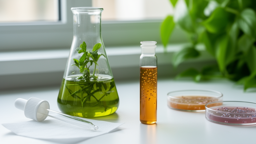
Catha edulis, an evergreen shrub of the Celastraceae family widely grown in East Africa and the Arabian Peninsula, brought its own chemical credentials to the project. Its young leaves are traditionally chewed for their stimulant effects, an activity attributed to amphetamine-like alkaloids, mainly cathinone and cathine. The leaves also contain flavonoids, tannins, glycosides, terpenoids, and phenolics, which the authors identify as natural reducing and stabilizing agents. For the synthesis, freshly purchased leaves from a local market in Bahir Dar were washed, air-dried, chopped, and heated in double-distilled water at 60 degrees Celsius for ten minutes. The filtered extract was stored at 4 degrees Celsius until use. In the core reaction, 50 milliliters of a 5 percent aqueous extract were combined with 50 milliliters of a 4 millimolar silver nitrate solution and left to react at room temperature, with the appearance of a deep red color providing the first visual sign that silver ions had been reduced to metallic silver.
Spectroscopy confirmed what the color change suggested. Ultraviolet-visible absorption spectroscopy revealed a distinct surface plasmon resonance band at 416 nanometers, a hallmark of silver nanoparticle formation that is consistent with previous reports on plant-extract-mediated synthesis. The team went further, applying a Tauc plot analysis to the absorption data under the assumption of a direct allowed electronic transition, and extrapolated the linear region near the absorption edge to obtain an effective optical transition energy of 2.65 electronvolts, a value the authors note agrees well with literature reports for silver nanoparticles. The analysis suggests that the optical response reflects a combination of nanoscale effects and electronic transitions associated with the particles, potentially influenced by the phytochemical components derived from the extract.
A substantial part of the study concerned optimization, because the concentration of extract, the reaction time, and the pH of the medium all shape how many particles form and how well they disperse. When different volumes of the 5 percent extract, from 10 to 100 milliliters, were mixed with a constant 50 milliliters of the 4 millimolar silver nitrate solution, low extract volumes gave weak surface plasmon resonance intensity, indicating insufficient reducing and stabilizing molecules, while the highest volume produced a red shift suggesting changes in particle size distribution, aggregation, or local surface environment. Based on the spectral characteristics, 75 milliliters of extract mixed with 50 milliliters of silver nitrate solution was identified as the optimum concentration for producing relatively stable nanoparticles.
Time and acidity mattered as well. Monitoring the reaction from 30 minutes to 72 hours showed the surface plasmon resonance intensity falling and shifting to longer wavelengths by 72 hours, which the authors interpret as nanoparticle aggregation and a decline in the concentration of well-dispersed particles; 48 hours was therefore identified as the optimal reaction time. Adjusting the pH with 0.1 molar hydrochloric acid or sodium hydroxide revealed a clear alkaline advantage: absorbance increased as the pH rose from 7 to 11, consistent with deprotonation of phytochemicals enhancing their reducing ability and promoting rapid nucleation of small, well-dispersed particles. Pushing the pH to 13 reversed the trend, decreasing the absorbance peak and producing a red shift attributed to aggregation. pH 11 was identified as the optimum condition for synthesis.
Structural characterization established the crystalline identity of the product. X-ray diffraction showed distinct peaks at 2-theta values of 38.23, 44.34, 64.52, and 77.39 degrees, indexed to the (111), (200), (220), and (311) planes of face-centered cubic metallic silver and matching the standard reference data in the Joint Committee on Powder Diffraction Standards database. The (111) reflection near 38.23 degrees was the most intense, indicating the predominant crystallographic orientation, and the sharp, well-defined peaks pointed to a high degree of crystallinity, while the absence of additional impurity peaks suggested the particles consist mainly of pure metallic silver. Applying the Debye-Scherrer equation to the diffraction data yielded an average crystallite size of 17.65 nanometers. The authors are careful to distinguish this coherently diffracting crystalline domain from overall particle size, which would require transmission or scanning electron microscopy to determine directly, although they note the two may be comparable for predominantly single-crystalline particles.
Infrared spectroscopy filled in the molecular picture. The leaf extract showed bands at 3411, 2329, 2078, 1638, 1075, and 588 inverse centimeters, while the nanoparticles showed bands at 3439, 2929, 2375, 1630, 1384, 1083, and 630 inverse centimeters. The broad bands near 3411 and 3439 inverse centimeters are attributed mainly to O-H stretching of hydroxyl-containing compounds such as phenols and alcohols, which may participate in silver ion reduction and nanoparticle stabilization. Bands associated with amide and aromatic vibrations, aliphatic C-H stretching, and C-O stretching of alcohols and ethers all point to organic phytochemicals on the particle surface. The shifts in major bands and changes in intensity between the extract and nanoparticle spectra support the involvement of hydroxyl, carbonyl, amine, and other oxygen-containing phytochemicals in reducing silver ions and capping the particles, although the authors caution that some individual band assignments, particularly at low frequency, cannot be conclusively established from FTIR alone.
Qualitative phytochemical screening of the extract independently confirmed the presence of flavonoids, tannins, phenols, glycosides, alkaloids, and steroids, identified through characteristic color changes and precipitate formation with standard reagents. These secondary metabolites are known to contribute to antioxidant, antimicrobial, and anti-inflammatory activities, and in the synthesis context they plausibly serve as the reducing and capping agents. The authors note, however, that the specific roles of individual phytochemicals were not individually confirmed in this study, an important caveat when interpreting the mechanistic claims.
The practical payoff came in the antibacterial assay. Using the disc diffusion method on Mueller-Hinton agar, with sterile discs impregnated with 50 microliters of the nanoparticle colloidal solution, gentamicin as a positive control, and sterile distilled water as a negative control, the team measured zones of inhibition after 24 hours of incubation at 37 degrees Celsius, with all assays performed in triplicate and reported as mean plus or minus standard deviation. The nanoparticles produced a zone of 21.20 plus or minus 0.78 millimeters against the Gram-negative bacterium Klebsiella pneumoniae and 18.63 plus or minus 0.53 millimeters against the Gram-positive Streptococcus pyogenes. The authors attribute the difference to cell wall architecture: Gram-negative bacteria possess a thin peptidoglycan layer and an outer membrane rich in lipopolysaccharides that may enhance nanoparticle interaction and penetration, whereas the thicker peptidoglycan of Gram-positive bacteria acts as a protective barrier. Prior studies cited in the paper indicate that once inside bacterial cells, silver nanoparticles can induce reactive oxygen species formation, damage membranes, disrupt DNA replication, and inhibit key enzymes. The authors recommend follow-up work including detailed particle size and morphology characterization and determination of minimum inhibitory and bactericidal concentrations before the particles’ antibacterial efficacy can be judged comprehensively.
Subject of Research: Green synthesis of silver nanoparticles from Catha edulis leaf extract and their antibacterial activity
Article Title: Optimized Catha edulis leaf extract mediated synthesis of silver nanoparticles and their antibacterial activity against selected pathogenic bacteria
Article References: Mamo, H. S., & Solomon, M. A. (2026). Optimized Catha edulis leaf extract mediated synthesis of silver nanoparticles and their antibacterial activity against selected pathogenic bacteria. Discover Metals, 59(1), Article 7. https://doi.org/10.1007/s13404-026-00377-1
Image Credits: AI Generated
DOI: 10.1007/s13404-026-00377-1
Keywords: silver nanoparticles, green synthesis, Catha edulis, antibacterial activity, X-ray diffraction, UV-Vis spectroscopy, FTIR, zone of inhibition, Klebsiella pneumoniae, Streptococcus pyogenes, nanotechnology, plant extract
Cite Scienmag News
Denise Maddox. (October 28, 2026). Khat Leaf Extract Yields Silver Nanoparticles That Inhibit Two Pathogenic Bacteria. Scienmag. https://scienmag.com/khat-leaf-extract-yields-silver-nanoparticles-that-inhibit-two-pathogenic-bacteria/
Denise Maddox. "Khat Leaf Extract Yields Silver Nanoparticles That Inhibit Two Pathogenic Bacteria." Scienmag, 28 October 2026, https://scienmag.com/khat-leaf-extract-yields-silver-nanoparticles-that-inhibit-two-pathogenic-bacteria/. Accessed 28 October 2026.
Denise Maddox. "Khat Leaf Extract Yields Silver Nanoparticles That Inhibit Two Pathogenic Bacteria." Scienmag. October 28, 2026. https://scienmag.com/khat-leaf-extract-yields-silver-nanoparticles-that-inhibit-two-pathogenic-bacteria/