


Every year, millions of patients undergo midline laparotomy, the workhorse incision of abdominal surgery that runs vertically along the midline of the belly. For a substantial fraction of them, the operation is only the beginning of the story. The repaired abdominal wall can gradually give way, allowing internal organs to bulge through the scar as an incisional hernia, a complication that brings pain, impaired body image, emergency reoperations and a heavy burden on health systems. A new prospective study from Spain, published in the Journal of Abdominal Wall Surgery, reports encouraging long-term results for a preventive strategy that remains stubbornly underused: reinforcing the healing incision with a synthetic mesh before any hernia has had a chance to form.
The research, known as the VISIBLE study, enrolled 180 adults across tertiary and secondary hospitals of the Spanish National Health Service between July 2016 and June 2022. Every participant carried more than one risk factor for incisional hernia, criteria that included age over sixty years, obesity, diabetes, chronic lung or heart disease, smoking, kidney or liver disease, immunosuppression, active cancer and emergency operations. The mean patient age was 64.6 years and the mean body mass index was 27.4 kilograms per square metre. Nearly three quarters of the procedures were performed for cancer, roughly one third were emergencies, and almost a quarter took place in contaminated or dirty surgical fields, settings in which the use of a prophylactic implant has traditionally been considered controversial.
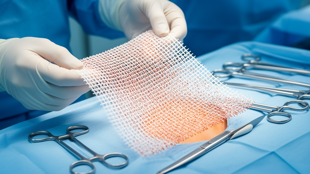
The technique at the heart of the trial was deliberately simple and reproducible. After closing the linea alba with a running long-term absorbable monofilament suture, the surgical team dissected a pocket in the prefascial plane, detached the umbilical scar from the midline, and laid a sheet of polyvinylidene fluoride, or PVDF, mesh over the repaired fascia with an overlap of at least two centimetres at each end. The mesh was fixed with absorbable stitches, the subcutaneous tissue was anchored to its surface to eliminate dead space, and a suction drain was left in place for three to five days. Because the mesh sits on top of the fascia rather than within the abdominal wall’s deeper muscular planes, the operation adds relatively little time and preserves the retromuscular space, a valuable reserve should a hernia ever need formal reconstruction later in life.
What makes the mesh genuinely novel is its chemistry. The implant, manufactured specifically for the study by FEG Textiltechnik in Aachen, Germany, is composed of 90 percent PVDF with a small fraction of triiron tetraoxide woven into its structure. Iron oxide is paramagnetic, which renders the otherwise hard-to-see implant clearly visible on magnetic resonance imaging. This ‘visible’ mesh allowed the investigators to track the implant’s dimensions, position and integrity directly over time, offering a window on mesh behaviour that physical examination alone cannot provide. Each mesh was 35 centimetres long and 7.5 centimetres wide, with a mean pore size of 2.6 millimetres and a weight of approximately 131 grams per square metre.
The results were striking. After two years of follow-up, which reached 96.1 percent of surviving patients, incisional hernia had developed in 21 patients in the intention-to-treat analysis, an incidence of 11.3 percent. When patients with early postoperative wound dehiscence were excluded in the per-protocol analysis, the figure fell to 8.9 percent. Given the clinical profile of the cohort, elderly oncology patients, emergency operations and contaminated fields included, the investigators consider these rates consistent with the lower end of published outcomes for high-risk laparotomy. Notably, three quarters of all hernias occurred among the first ninety patients recruited, hinting that surgical teams became more adept at selecting and managing patients as the study progressed.
Magnetic resonance imaging provided some of the most technically revealing data. MRI was performed in 78.1 percent of patients at six weeks and 65.1 percent at one year, and one hundred patients underwent imaging at both time points. Comparison of the measurements showed that the mesh neither shrank nor distorted significantly: mean length changed from 20.3 to 19.9 centimetres and mean width from 7.02 to 7.04 centimetres, differences that were not statistically significant. Mesh contraction, a well-recognised phenomenon with some synthetic implants that can expose unfilled gaps to rising intra-abdominal pressure, appears to be absent with this material, an observation the authors describe as an important strength of the MRI-based assessment.
Complications were, on the whole, manageable. Surgical-site infection occurred in 12.8 percent of patients during the index admission and seroma in 9.4 percent, although MRI detection pushed the seroma rate to 20.7 percent at six weeks, most of these fluid collections being asymptomatic and invisible on clinical examination. By three months, only 2.4 percent of patients still had a seroma. Mesh removal was ultimately required in four patients, just 2.2 percent of the cohort. Pain, often cited as a fear with mesh implants, was negligible: mean laparotomy pain on a visual analogue scale was 0.2 at three months, and no patient required long-term analgesia or mesh removal for chronic pain. Crucially, emergency surgery and contaminated fields were not associated with a higher rate of mesh-related complications, countering one of the strongest objections to prophylactic implantation.
The statistical modelling identified two independent predictors of hernia formation. Each additional unit of body mass index raised the odds by 19 percent, while patients who required a stoma faced nearly six-fold higher odds, with cumulative incidence at two years of about 27 percent for those with an ileostomy or colostomy compared with 6.3 percent for patients without one. Surprisingly, longer operative time was associated with slightly lower hernia risk, a counterintuitive finding the authors urge readers to treat cautiously, given the observational design and the possibility of residual confounding. Surgical-site infection showed a trend toward increased hernia risk that did not reach statistical significance, and all deaths in the cohort occurred among cancer patients, none related to the mesh.
The study does carry limitations that its authors acknowledge candidly. There was no randomised control group, one hospital contributed nearly 43 percent of patients and 75 percent of hernia events, the multivariable model was built on a modest number of outcome events, and the trial, partly conducted during the COVID-19 pandemic, lacked a standardised fascial closure protocol and validated patient-reported quality-of-life measures. The investigators used Firth penalised regression and bootstrap internal validation to guard against small-sample bias, but they emphasise that the findings are hypothesis-generating and must be confirmed in controlled comparative studies before prophylactic mesh becomes standard practice.
Yet the gap the study addresses is real and persistent. Surveys suggest that while roughly 85 percent of surgeons recognise the benefit of prophylactic mesh reinforcement, only 3 to 15 percent use it routinely, and up to half of patients express concerns about receiving a mesh without an existing hernia. By demonstrating that a simple onlay technique, applied by general surgeons rather than abdominal wall specialists across hospitals of varying size, can achieve a low hernia rate with acceptable morbidity and no significant mesh contraction on MRI, the VISIBLE study makes a persuasive case that the barrier is cultural as much as technical. If larger controlled trials bear out these results, the routine question at the end of high-risk laparotomy may shift from whether to place a mesh to why any surgeon would leave the healing incision unprotected.
Subject of Research: Prophylactic onlay PVDF mesh reinforcement for prevention of incisional hernia after high-risk midline laparotomy
Article Title: Incisional hernia prevention in high-risk midline laparotomy using onlay PVDF mesh: multicenter prospective observational study
Article References: Incisional hernia prevention in high-risk midline laparotomy using onlay PVDF mesh: multicenter prospective observational study. (n.d.). https://doi.org/10.3389/jaws.2026.17201
Image Credits: AI Generated
Keywords: incisional hernia, midline laparotomy, prophylactic mesh, PVDF, MRI visibility, emergency surgery, contaminated surgical field, abdominal wall closure, stoma, surgical site infection, surgical outcomes, Spain
Cite Scienmag News
Ophelia Keating. (October 15, 2026). Iron-Loaded Surgical Mesh Offers New Hope Against Hernias After High-Risk Abdominal Surgery. Scienmag. https://scienmag.com/iron-loaded-surgical-mesh-offers-new-hope-against-hernias-after-high-risk-abdominal-surgery/
Ophelia Keating. "Iron-Loaded Surgical Mesh Offers New Hope Against Hernias After High-Risk Abdominal Surgery." Scienmag, 15 October 2026, https://scienmag.com/iron-loaded-surgical-mesh-offers-new-hope-against-hernias-after-high-risk-abdominal-surgery/. Accessed 15 October 2026.
Ophelia Keating. "Iron-Loaded Surgical Mesh Offers New Hope Against Hernias After High-Risk Abdominal Surgery." Scienmag. October 15, 2026. https://scienmag.com/iron-loaded-surgical-mesh-offers-new-hope-against-hernias-after-high-risk-abdominal-surgery/


