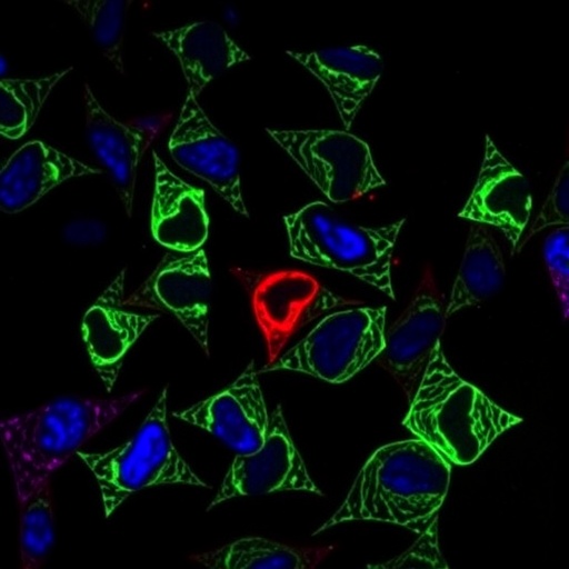
In a groundbreaking study poised to reshape our understanding of pathological scarring, researchers have unveiled a novel mechanism by which interferon-gamma (IFN-γ) induces ferroptosis in keloid fibroblasts, unlocking exciting therapeutic possibilities for refractory keloid disorders. Published in the prestigious journal Cell Death Discovery, this investigation illuminates the pivotal role of IFN-γ in regulating ferroptotic cell death through suppression of serpine2 expression—an insight that not only deepens scientific comprehension but also offers a promising avenue to mitigate the relentless progression of keloids.
Keloids, characterized by excessive fibroblast proliferation and extracellular matrix deposition beyond original wound boundaries, persist as an enigmatic clinical challenge, often resisting conventional therapies. Traditional interventions—ranging from corticosteroid injections to surgical excision—frequently yield inconsistent outcomes, stimulating a fervent search for molecular targets that can selectively modulate fibroblast viability. The emergence of ferroptosis, a regulated form of iron-dependent cell death distinguished by lipid peroxidation, has introduced a new dimension to cell fate regulation, distinct from apoptosis or necrosis, with profound implications in various pathologies. This recent study pinpoints IFN-γ as a critical instigator of ferroptotic demise in the notoriously resilient keloid fibroblasts.
The authors, Huang, Yu, Luo, and colleagues, meticulously demonstrated that exposure of keloid-derived fibroblasts to IFN-γ significantly curtails the expression of serpine2, a serine protease inhibitor previously implicated in extracellular matrix regulation and cellular survival pathways. This downregulation of serpine2 acts as a molecular switch that sensitizes fibroblasts to ferroptosis by unleashing uncontrolled lipid peroxidation and iron-dependent reactive oxygen species accumulation. These findings underscore serpine2’s heretofore underappreciated function as a ferroptosis gatekeeper within fibrotic contexts, adding a nuanced perspective to its biological repertoire.
Through a series of precisely controlled in vitro experiments, the research team utilized ferroptosis-specific inhibitors and genetic modulation techniques to validate that the cytotoxic effects of IFN-γ on keloid fibroblasts derive not from canonical apoptotic cascades but rather from ferroptotic pathways. Lipid peroxidation assays revealed striking elevations in malondialdehyde and 4-hydroxynonenal levels, hallmark indicators of ferroptotic damage, concurrent with diminished glutathione peroxidase 4 (GPX4) activity—a vital antioxidant enzyme counteracting lipid peroxidation. Importantly, restoration of serpine2 expression was able to partially rescue fibroblasts from IFN-γ-induced ferroptosis, cementing its functional importance.
These revelations bear profound clinical weight, as keloid pathology involves sustained fibroblast activation and extracellular remodeling that empower excessive scar tissue formation. By harnessing IFN-γ’s ability to incapacitate keloid fibroblasts via ferroptosis induction, it becomes conceivable to strategically target keloid lesions at the cellular level. This paradigm shift elevates ferroptosis from a mere biochemical curiosity to a therapeutic mechanism with tangible translational potential. Given the historical difficulty in controlling fibroblast proliferation without collateral tissue damage, a ferroptosis-driven approach could enable highly selective disruption of pathological cells while preserving normal skin architecture.
Further molecular dissections revealed that IFN-γ signaling orchestrates a multi-tiered suppression of serpine2 at the transcriptional level, likely mediated by STAT1-driven modulation of promoter accessibility. This intricate regulation highlights the convergence of immune cytokine signaling and metabolic stress pathways in dictating fibroblast fate decisions. The intersection of chronic inflammation and ferroptosis provides a compelling framework for future exploration, particularly in the context of other fibrotic diseases where maladaptive tissue remodeling features prominently.
Intriguingly, the study also observed that IFN-γ treatment led to alterations in intracellular iron homeostasis, potentiating ferroptotic susceptibility. Elevated expression of transferrin receptor and decreased ferritin levels signified a remodeling of cellular iron flux, fueling the iron-dependent lipid peroxidation central to ferroptosis. This iron dysregulation, compounded by serpine2 inhibition, acts synergistically to drive fibroblasts toward a ferroptotic endpoint. Such multifaceted regulation underscores the complex interplay between cytokines, iron metabolism, and cell death modalities in pathological fibrosis.
From a therapeutic development perspective, these findings pave the way for innovative interventions that could exploit IFN-γ or its downstream effectors to induce targeted ferroptosis in keloid fibroblasts. Drug candidates designed to mimic or amplify IFN-γ’s ferroptotic capacity, combined with iron chelators or lipid peroxidation enhancers, could form a rational combination to effectively ablate stubborn keloid scars. Preclinical models will be essential to refine dosing strategies and minimize off-target effects, ensuring safety and specificity in delicate dermal tissues.
The ramifications extend beyond dermatology, as ferroptosis is implicated in oncological, neurological, and cardiovascular diseases. Understanding how immune factors like IFN-γ interface with ferroptotic machinery could unlock approaches to selectively eliminate pathogenic cell populations across a spectrum of fibrotic and neoplastic conditions. This convergence of immunology and ferroptosis biology heralds a new era of targeted molecular therapies aimed at modulating cellular ferroptotic thresholds in disease settings.
While the clinical translation remains in early stages, the clarity provided by Huang and colleagues’ work offers a compelling blueprint for moving forward. It challenges existing paradigms by positioning IFN-γ not merely as an inflammatory cytokine but as a critical modulator of ferroptotic cell fate in keloid fibroblasts, unveiling therapeutic vulnerabilities that were previously untapped. The balance between immune modulation, metabolic stress, and ferroptosis may well redefine strategies for intractable fibrotic diseases.
As further research elucidates the detailed signaling networks and metabolic parameters governing ferroptosis in fibroblasts from diverse origins, tailored therapies could emerge that harness or temper IFN-γ activity to beneficial ends. Precision medicine approaches might integrate patient-specific fibrosis profiles, cytokine milieu assessments, and ferroptotic susceptibility markers to customize interventions with maximal efficacy and minimal adverse effects.
This study also prompts reconsideration of the broader implications of inflammation-fueled ferroptosis in tissue homeostasis and pathology. Chronic inflammatory environments characterized by elevated IFN-γ levels—typical in autoimmune and infectious dermatoses—could inadvertently provoke ferroptotic tissue damage or remodeling, influencing disease progression. Dissecting these complex interactions will be vital for developing balanced therapies that restore tissue integrity without exacerbating injury.
Collectively, the identification of IFN-γ as a potent ferroptosis inducer through serpine2 inhibition in keloid fibroblasts heralds a transformative advance in fibrosis research. Beyond elucidating fundamental disease mechanisms, it charts a course toward viable, mechanistically grounded treatments for patients plagued by recalcitrant keloid scars. The fusion of immunological insights with ferroptosis biology exemplifies the forefront of translational medicine, offering hope for improved outcomes in an area long marred by therapeutic limitations.
In summary, this landmark investigation expands the scientific landscape by establishing a direct causal link between IFN-γ signaling and ferroptotic cell death in keloid fibroblasts via repression of serpine2. The ramifications span fundamental biology, disease pathogenesis, and therapeutic innovation, positioning ferroptosis induction as a promising strategy in combatting pathological skin fibrosis. As the global burden of keloid scarring persists, this research infuses renewed optimism, driving the scientific community toward novel, targeted solutions.
Subject of Research: IFN-γ-induced ferroptosis in keloid fibroblasts via inhibition of serpine2 expression
Article Title: IFN-γ could induce ferroptosis in keloid fibroblasts by inhibiting the expression of serpine2
Article References: Huang, J., Yu, S., Luo, J. et al. IFN-γ could induce ferroptosis in keloid fibroblasts by inhibiting the expression of serpine2. Cell Death Discov. 11, 217 (2025). https://doi.org/10.1038/s41420-025-02401-3
Image Credits: AI Generated


