


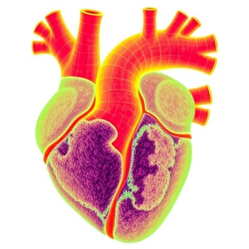
Mitral valve prolapse, a condition in which the valve between the heart’s left atrium and left ventricle bulges backward into the atrium during each heartbeat, affects millions of people worldwide and accounts for a substantial share of the roughly 40,000 deaths attributed to non-rheumatic primary mitral valve disease each year. When the condition produces moderate to severe mitral regurgitation—the backflow of blood that can culminate in heart failure, arrhythmia, and death—the only current remedy is surgery, either repair or replacement of the valve. Yet even after a technically successful operation, adverse post-operative remodeling of the valve tissue can drive recurrent leakage, forcing some patients back onto the operating table. Now, a team of researchers working in the field of cardiac mechanobiology has uncovered a fundamental difference in how the cells within prolapsed valves behave at the most basic mechanical level, a finding that may explain why some repaired valves fail and could point the way toward the first non-surgical therapies for the disease.
The study, published in Annals of Biomedical Engineering, focused on mitral valve interstitial cells, or MVICs, the resident cells embedded within the collagen-rich extracellular matrix that gives the valve leaflet its remarkable combination of strength and flexibility. In healthy tissue, these cells quietly maintain the matrix, synthesizing and degrading structural proteins as needed to keep the leaflet functioning across billions of cardiac cycles. In disease, however, MVICs can adopt an activated phenotype, characterized by elevated expression of the contractile protein alpha-smooth muscle actin, along with excess collagen deposition, matrix fragmentation, and increased activity of matrix metalloproteinases, the enzymes that break down structural proteins. The result is a valve leaflet that thickens and stiffens, further distorting the delicate mechanics that keep it closing properly. What remained unknown until now is whether these altered cellular behaviors are intrinsic to the cells themselves, or merely a response to an abnormal tissue environment—and precisely how they change the mechanical landscape around each cell.
To answer that question, the researchers, led by Toni M. West and Micheal S. Sacks, isolated MVICs from three patients undergoing first-time surgery for mitral valve prolapse and from three donors whose valves were anatomically normal. Rather than studying the cells in flat culture dishes or in opaque excised tissue—both of which obscure the true three-dimensional mechanical dialogue between a cell and its surroundings—the team embedded the isolated cells in engineered hydrogels designed to mimic key aspects of the native cellular microenvironment. The gels were built from eight-arm poly(ethylene) glycol functionalized with norbornene groups, crosslinked with peptide sequences that cells can enzymatically degrade, and decorated with CRGDS adhesion peptides that allow cells to grip their surroundings. Suspended within each gel were fluorescent microspheres roughly one micrometer in diameter, serving as fiducial markers whose movements would betray every tug and push exerted by the encapsulated cells.
After three days of culture, each hydrogel was imaged twice on a laser scanning confocal microscope: once in the basal state, with the cells actively contracting, and once in a fully relaxed state induced by cytochalasin-D, a drug that dismantles the actin cytoskeleton. By subtracting the relaxed configuration from the contracted one, the researchers obtained precise three-dimensional displacement fields for thousands of microspheres surrounding each cell, captured at an isotropic voxel resolution of just 0.288 micrometers. The key innovation of the study, however, lay not in the imaging but in the computational machinery applied to those images. Conventional traction force microscopy, a technique dating back to 1999, assumes that the hydrogel surrounding a cell remains mechanically uniform, an assumption that is demonstrably false when cells are actively degrading and rebuilding their matrix. The new study deployed a recently developed framework that dispenses with that assumption entirely.
In the inverse modeling approach, the hydrogel was treated as a compressible Neo-Hookean hyperelastic solid, an appropriate description given the significant compressibility of poly(ethylene) glycol gels, which must be accounted for to recover accurate mechanical properties. The local stiffness of the gel was encoded as a spatially varying material parameter expressed in exponential form, allowing the model to capture changes in modulus spanning large orders of magnitude while remaining numerically stable. The team then solved a nonlinear optimization problem: using the limited-memory Broyden–Fletcher–Goldfarb–Shannon algorithm, they iteratively adjusted the simulated stiffness field until the computed displacements matched the experimentally measured ones throughout a reliable measurement zone the authors call the “event horizon,” defined by a displacement cutoff of 0.38 micrometers corresponding to three standard deviations of imaging noise. Gradient computations performed with adjoint methods and Tikhonov regularization kept the ill-posed inverse problem stable, and convergence was reached in roughly an hour per hydrogel on a 24-core desktop workstation. The output was a full three-dimensional map of how each cell had locally stiffened or softened its surroundings, from which surface traction forces and local strain energy densities could be computed using the first Piola–Kirchhoff stress tensor.
The results delivered a surprise on multiple fronts. Collagen staining confirmed that cells deposited collagen in their immediate vicinity, producing pronounced local stiffening close to the cell membrane, while regions farther from the cell surface actually softened, reflecting enzymatic degradation of the gel’s degradable crosslinkers. Cells from prolapsed valves stiffened their gels more and degraded them less than cells from normal valves, consistent with the known tendency of diseased valve cells to overproduce collagen. But the most striking finding concerned contractility. The prolapsed-valve cells exhibited larger basal contractile displacements, squeezing their surroundings more deeply, yet the actual traction forces they generated and the strain energy they injected into the gel were significantly lower than those of normal cells. In other words, the diseased cells appeared to be working harder visually while doing less mechanical work—an apparent paradox that only became visible because the model accounted for the cells’ remodeling of their own microenvironment.
The interpretation is subtle but consequential. Because the cells from prolapsed valves had remodeled their surroundings so extensively, depositing stiffer matrix nearby, they could achieve large displacements with comparatively modest force. A conventional analysis assuming a uniform gel would have misread this picture entirely, potentially overestimating the force output of the diseased cells. By capturing the true, heterogeneous mechanical state of the gel, the new framework revealed that prolapsed-valve interstitial cells are not simply hypercontractile muscle-like cells, as their elevated alpha-smooth muscle actin expression might suggest, but cells with a fundamentally rebalanced mechanical portfolio: more matrix deposition, less degradation, deeper but weaker contractions.
Crucially, because the experiments were performed on isolated cells in standardized gels, the observed differences cannot be attributed to the altered biochemical environment of a diseased valve. The behaviors are intrinsic to the cell phenotype itself, apparently persisting even after the cells were removed from the patient’s tissue and cultured at low passage on collagen-coated dishes designed to prevent artificial activation. This intrinsic quality carries a sobering implication for surgeons and patients alike: repairing the geometry of a prolapsed valve does not necessarily correct the underlying cellular behavior that drove the disease, and those intrinsic behaviors may continue remodeling the repaired tissue in ways that promote recurrent regurgitation and secondary surgery.
The study’s authors suggest that this new mechanobiological window could reshape how post-repair remodeling is understood and, eventually, treated. If the contractile and matrix-remodeling behaviors of valve interstitial cells can be measured, perhaps from cells harvested during surgery, clinicians might one day stratify patients by risk of recurrent disease, identifying whose valves are likely to remodel adversely after repair. More ambitiously, drugs that modulate the contractility or matrix-synthesis programs of these cells could be developed as adjuncts to surgery or even as alternatives, targeting the cellular engines of the disease rather than its structural consequences. Such therapies have remained elusive precisely because the cellular mechanisms of post-repair remodeling were so poorly characterized; this work supplies the first direct, quantitative evidence that prolapsed-valve cells behave mechanically differently from normal ones in ways that could be pharmacologically targeted.
The technical achievement also extends beyond the mitral valve. The inverse modeling framework, which simultaneously recovers cellular tractions and cell-induced changes in local material properties for compressible, degradable hydrogels, offers a template for studying any cell type whose remodeling behavior complicates conventional traction measurements, including cancer cells, fibroblasts in fibrotic disease, and cells in engineered tissues. For a condition that has historically been managed exclusively with scalpels and sutures, the demonstration that its cellular drivers can be measured, quantified, and distinguished from healthy behavior marks a meaningful step toward understanding—and perhaps one day preventing—the silent remodeling that undermines valve repair. As the researchers conclude, the behaviors of mitral valve interstitial cells may determine how prolapsed valves respond to repair, and for the first time, those behaviors can now be seen with clarity.
Cite Scienmag News
Ophelia Keating. (September 6, 2026). Human Mitral Valve Cells Show Distinct 3D Remodeling in Prolapse. Scienmag. https://scienmag.com/human-mitral-valve-cells-show-distinct-3d-remodeling-in-prolapse/
Ophelia Keating. "Human Mitral Valve Cells Show Distinct 3D Remodeling in Prolapse." Scienmag, 6 September 2026, https://scienmag.com/human-mitral-valve-cells-show-distinct-3d-remodeling-in-prolapse/. Accessed 6 September 2026.
Ophelia Keating. "Human Mitral Valve Cells Show Distinct 3D Remodeling in Prolapse." Scienmag. September 6, 2026. https://scienmag.com/human-mitral-valve-cells-show-distinct-3d-remodeling-in-prolapse/


