


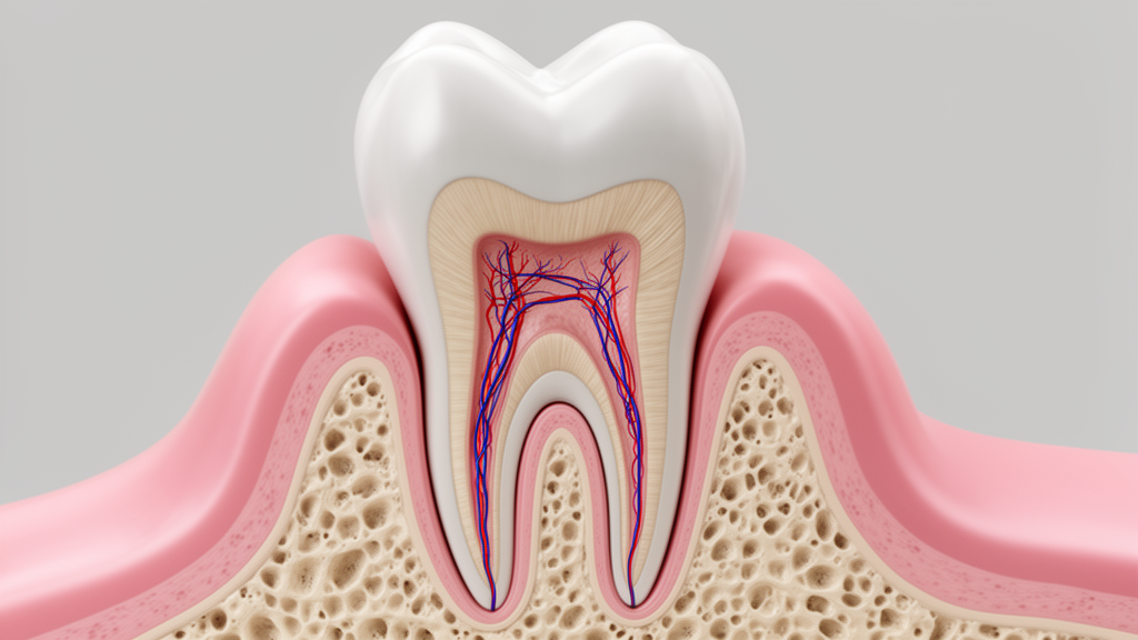
In a remarkable demonstration of the body’s innate healing capacity, clinicians have documented the successful regeneration of a child’s severely damaged molar tooth using cutting-edge biological techniques rather than traditional root canal procedures. The case, published in Clinical Case Reports, details how a 9-year-old girl’s immature mandibular first molar—infected, painful, and structurally compromised—was restored to health through a regenerative endodontic procedure that coaxed the tooth’s own stem cells into rebuilding lost tissue. The results, tracked over twelve months, offer compelling evidence that teeth once destined for extraction or brittle apexification treatments can instead be guided toward genuine biological recovery.
The patient arrived with localized pain in her lower right first molar, a tooth that had previously been restored with amalgam and showed classic signs of deep infection: tenderness on percussion and an extraoral sinus tract—a small drainage channel visible on the outside of her chin that signaled chronic apical abscess. Radiographic imaging revealed the full extent of the problem. The tooth’s roots were still developing, with wide-open apices and dangerously thin dentinal walls, accompanied by a distinct radiolucency around the root tips indicating active periapical infection. In a tooth this young and underdeveloped, conventional root canal treatment posed serious long-term risks, as the structural integrity of the root would remain permanently compromised.
Historically, dentists facing necrotic immature permanent teeth have relied on apexification—using calcium hydroxide or mineral trioxide aggregate (MTA) to stimulate apical closure. While these methods can seal the root tip, they leave the tooth structurally flawed: the dentinal walls never thicken, the root never lengthens, and the resulting tooth remains vulnerable to fracture under normal chewing forces. Regenerative endodontic procedures (REPs) represent a fundamentally different philosophy. Rather than simply sealing off the infection, REPs aim to harness the tooth’s own biological machinery—specifically stem cells from the apical papilla (SCAP)—to rebuild living tissue inside the canal space and encourage continued root maturation.
The conceptual foundation of REP rests on the classic triad of tissue engineering: stem cells, scaffolds, and signaling molecules. By deliberately inducing bleeding from the periapical tissues into the cleaned canal space, clinicians introduce a rich population of SCAP alongside a natural blood clot scaffold loaded with endogenous growth factors. These elements work in concert to facilitate healing and tissue regeneration from within. The approach demands a delicate balance: the canal must be thoroughly disinfected to eliminate harmful bacteria, yet the treatment must preserve the viability of the very stem cells needed for regeneration. This is achieved through minimal mechanical instrumentation and passive irrigation with biocompatible solutions such as low-concentration sodium hypochlorite and 17% EDTA, which conditions the dentin surface and facilitates the release of growth factors embedded in the tooth structure.
In this case, the treatment protocol was executed across two carefully sequenced visits. During the first appointment, following inferior alveolar nerve block anesthesia with 1.8 mL of 2% lidocaine containing epinephrine, the tooth was isolated with a rubber dam and accessed. Mechanical instrumentation was deliberately kept to a minimum—using only ISO #30 and #40 K-files—to avoid disrupting the apical stem cell niche. The canals were irrigated with 1.5% sodium hypochlorite followed by saline, and a double antibiotic paste composed of ciprofloxacin and metronidazole was placed as an intracanal medicament before temporary sealing. Three weeks later, at the second visit, anesthesia was achieved using 3% mepivacaine without a vasoconstrictor—a deliberate choice, as vasoconstrictors would suppress the bleeding needed for scaffold formation. The antibiotic paste was removed with minimal instrumentation and saline irrigation, followed by a 17% EDTA rinse to condition the dentin and release growth factors.
The pivotal moment came when intentional bleeding was induced by gently over-instrumenting approximately 1 to 2 millimeters beyond the apex with a sterile #25 K-file. This triggered the formation of a blood clot within the canal—a natural scaffold rich in growth factors and stem cells that would serve as the biological foundation for regeneration. Mineral trioxide aggregate was then placed approximately 3 millimeters below the cementoenamel junction to provide a biocompatible coronal seal, confirmed radiographically. A moist cotton pellet protected the MTA, the cavity was sealed temporarily, and the patient was referred for a permanent amalgam restoration to ensure a reliable coronal seal given the extensive loss of coronal tooth structure.
At the twelve-month follow-up, the results were striking. The patient was completely asymptomatic, and the extraoral sinus tract had fully resolved. Radiographs demonstrated healing of the periradicular lesion, continued root maturation with visibly increased dentinal wall thickness, and clear evidence of apical closure. Clinical examination confirmed soft tissue healing, and pulp sensitivity tests documented progressive recovery across multiple assessment points. These outcomes represent exactly what regenerative endodontics aims to achieve: not merely the elimination of infection, but the genuine biological restoration of tooth structure that conventional apexification cannot provide.
Several factors likely contributed to this success. The patient’s young age—just 9 years old—is consistently recognized as one of the most important predictors of regenerative outcomes, as younger patients exhibit higher concentrations of viable SCAP and enhanced angiogenic potential. Research has shown that stem cells from the apical papilla retain high viability and migratory potential even when exposed to calcium silicate-based materials, and they respond robustly to growth factors such as VEGF, PDGF, and TGF-β1. A large-scale retrospective study by Jiang and Liu confirmed that younger age at treatment was significantly associated with achieving both primary and secondary regenerative outcomes. The open apex in this case also facilitated stem cell influx and sufficient intracanal bleeding, with studies reporting a mean reduction of 56.8% in foramen size following REP and complete apical closure in 26.7% of immature teeth treated, with best outcomes in patients younger than 12.
Notably, posterior teeth like this molar may actually hold an advantage in regeneration despite their anatomical complexity. Recent studies have demonstrated that molars exhibit superior regenerative outcomes compared to anterior teeth, with a mean increase of 39% in root area and 11% in root length for posterior teeth, versus 26% and 6% respectively for anterior teeth. However, the multirooted structure and curved canal systems of molars pose considerable challenges for disinfection and uniform bleeding induction across all canals. The choice of a double antibiotic paste over the more commonly used triple antibiotic paste was also strategic—it eliminated the risk of coronal discoloration associated with minocycline while maintaining effective antimicrobial activity, a finding supported by comparative research showing equal efficacy of DAP for root maturation and apical healing.
While this case demonstrates the remarkable potential of regenerative endodontics, the authors acknowledge important limitations. The twelve-month follow-up represents short-term observation, and longer-term studies have shown that regenerative treatments can initially succeed yet still develop complications such as recurrent apical periodontitis several years later. As a single case report, the findings cannot be generalized without larger clinical trials, and histologic evaluation of the newly formed tissue was not possible in a clinical setting. Nevertheless, the case adds to a growing body of evidence that regenerative endodontic procedures—when applied with careful case selection, effective disinfection, deliberate bleeding induction, and a durable coronal seal—offer a biologically superior alternative to conventional apexification, preserving natural dentition and supporting genuine root development in young patients whose teeth still hold regenerative promise.
Subject of Research: Regenerative endodontic treatment of a necrotic immature mandibular first molar in a child
Article Title: Regenerative Endodontic Procedure in Mandibular First Molar: A Clinical Case Report
Article References: Saeedi, S., Hamedi, A., Mansouri, M., & Shiezadeh, I. (2026). Regenerative Endodontic Procedure in Mandibular First Molar: A Clinical Case Report. Clinical Case Reports, 14(10), Article e73610. https://doi.org/10.1002/ccr3.73610
Image Credits: AI Generated
DOI: 10.1002/ccr3.73610
Keywords: regenerative endodontics, dental pulp regeneration, stem cells from the apical papilla, immature permanent teeth, apexification, mineral trioxide aggregate, double antibiotic paste, mandibular first molar, root maturation, pediatric dentistry, blood clot scaffold, case report
Cite Scienmag News
Ophelia Keating. (October 17, 2026). How a 9-Year-Old’s Molar Regrew Its Own Root: Inside a Landmark Regenerative Dental Case. Scienmag. https://scienmag.com/how-a-9-year-olds-molar-regrew-its-own-root-inside-a-landmark-regenerative-dental-case/
Ophelia Keating. "How a 9-Year-Old’s Molar Regrew Its Own Root: Inside a Landmark Regenerative Dental Case." Scienmag, 17 October 2026, https://scienmag.com/how-a-9-year-olds-molar-regrew-its-own-root-inside-a-landmark-regenerative-dental-case/. Accessed 17 October 2026.
Ophelia Keating. "How a 9-Year-Old’s Molar Regrew Its Own Root: Inside a Landmark Regenerative Dental Case." Scienmag. October 17, 2026. https://scienmag.com/how-a-9-year-olds-molar-regrew-its-own-root-inside-a-landmark-regenerative-dental-case/


