


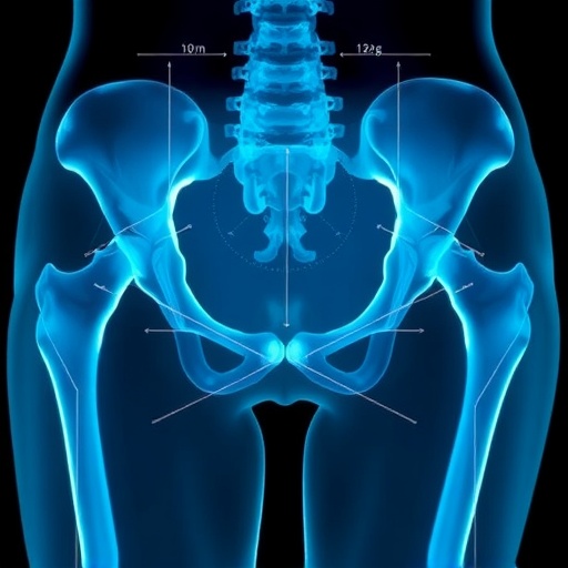
A routine hip X-ray may contain a surprisingly clear warning sign for osteoporosis, even when the condition has never been diagnosed. In a study of women preparing for total hip arthroplasty, researchers found that measuring the width of the bone’s inner canal at a specific point on an ordinary anteroposterior radiograph could identify patients likely to have severely reduced bone mineral density. The most practical measurement, called the canal–bone ratio at 7 centimeters below the lesser trochanter, or CBR7, performed nearly as well as more complicated radiographic calculations. The finding could give orthopedic surgeons an inexpensive way to flag patients who need formal testing before or after surgery, potentially helping prevent fractures around a hip implant.
The study, led by researchers at Osaka University and collaborating institutions in Japan, examined 317 women who underwent primary total hip arthroplasty. All had preoperative bone mineral density measured with dual-energy X-ray absorptiometry, commonly known as DXA, on the side scheduled for surgery. DXA remains the standard method for diagnosing osteoporosis because it estimates bone mineral density and produces a T-score relative to a healthy young-adult reference population. Under the study’s criteria, osteopenia was defined as a T-score between –1 and –2.5, while osteoporosis was defined as a T-score of –2.5 or lower. The investigators then compared those results with measurements extracted from the patients’ existing hip radiographs.
The clinical problem is substantial. Osteoporosis weakens the internal architecture of bone, reducing its ability to withstand ordinary forces and increasing the risk of fractures during or after joint replacement. A fragile femur can complicate the insertion and fixation of an implant, while weakened bone may be more vulnerable to fractures around the prosthesis later. Yet patients undergoing hip replacement do not always receive DXA testing, partly because of cost, limited availability, scheduling constraints, or the assumption that their orthopedic problem is separate from systemic bone loss. In the Osaka cohort, osteoporosis was detected in 129 women, or 40.7 percent of the sample. Only 48 of those patients, representing 37.2 percent, were receiving osteoporosis treatment.
The treatment gap was even wider when partially treated patients were included. Twenty-four women were taking osteoporosis medication but still had T-scores at or below –2.5, placing them in the study’s “undertreated” category. Together, untreated and undertreated patients accounted for 105 of the 129 women with osteoporosis, or 81.4 percent. The authors caution that their estimate may actually be conservative because they classified patients using the lowest T-score from the hip undergoing surgery rather than the lowest value across all relevant skeletal sites, such as the lumbar spine and opposite hip. Even so, the results show why the preoperative period may provide an important opportunity to detect bone disease that otherwise remains invisible.
To search for a useful radiographic signal, the researchers evaluated eight indices on digitally stored anteroposterior hip images. Some describe the shape of the upper femur, while others quantify how much of the bone’s width or area is occupied by the medullary canal—the hollow, marrow-containing space inside the shaft. The canal–bone ratio is calculated by dividing the canal width by the total width of the femur at a specified level. The team measured this ratio 2, 7, and 10 centimeters below the lesser trochanter, a bony landmark near the top of the femur. They also calculated canal–bone area ratios across segments extending from 2 to 7, 2 to 10, and 7 to 10 centimeters below the landmark. A wider canal relative to the entire bone suggests thinning of the surrounding cortical shell, a structural change associated with loss of bone strength.
The investigators assessed each index using receiver operating characteristic analysis, a statistical method that measures how well a test distinguishes between two conditions. Its central summary is the area under the curve, or AUC. An AUC of 0.5 indicates performance no better than chance, whereas an AUC of 1.0 represents perfect discrimination. The strongest result came from the canal–bone area ratio measured between 2 and 7 centimeters below the lesser trochanter, which achieved an AUC of 0.801. The area ratio from 7 to 10 centimeters and the single-point CBR7 each produced an AUC of 0.798. The remaining values were lower: 0.785 for the 2-to-10-centimeter area ratio, 0.766 for CBR10, 0.740 for CBR2, 0.696 for the canal flare index, and 0.654 for the morphological cortical index.
Although the area-based measurements were marginally strongest, CBR7 offers an important practical advantage: it requires only one width measurement at one anatomical level. The researchers found that a CBR7 cutoff of approximately 0.51 could provide a highly sensitive screen, meaning that the measurement could identify more than 90 percent of patients with osteoporosis when the threshold was selected to favor sensitivity. In simple terms, a canal wider than roughly half the total femoral width at that location could signal the need for DXA evaluation. This threshold is not a diagnosis, and it would likely produce some false-positive results, but it could help surgeons decide which patients should receive confirmatory testing.
The measurement also appeared relatively robust across different clinical circumstances. Many women in the primary cohort had osteoarthritis associated with developmental dysplasia of the hip, a condition that changes the shape and mechanics of the joint. Although diagnostic performance tended to be somewhat lower in that group than among patients with other hip diseases, the cutoff values for the canal-based indices differed little between groups. CBR7 showed the smallest absolute difference, just 0.007. The researchers further tested the approach in an independent cohort from another institution. Of 209 consecutive women undergoing hip replacement, 148 remained after applying the study’s exclusion criteria, and the canal-based indices retained similar cutoff values, with differences for the CBR measurements of less than 0.009.
Reliability is crucial for any tool intended for routine clinical use, because a measurement that changes substantially between observers would be difficult to trust. In repeated assessments, the study found that CBR7 and the canal–bone area ratios had strong reproducibility. Interobserver intraclass correlation coefficients, which quantify agreement between different measurers, ranged from 0.560 to 0.882 across all indices, while intraobserver values ranged from 0.827 to 0.952 when the same observer repeated the measurements. The most reliable measures were among the area ratios, but CBR7 combined high performance with a smaller measurement burden and a greater likelihood of being visible on standard hip radiographs. Images do not always extend far enough down the femur to include the 10-centimeter level, making the 7-centimeter location more broadly applicable.
The study does not suggest that an X-ray can replace DXA, nor that every person with a wide-appearing femoral canal has osteoporosis. A plain radiograph is a two-dimensional projection, and its measurements can be affected by positioning, magnification, image quality, and variations in femoral anatomy. The researchers controlled magnification using a metal sphere included in the imaging protocol and standardized leg rotation, but real-world images may be less consistent. Their work was also retrospective and centered on Japanese women undergoing hip replacement at a limited number of institutions. Bone shape differs with sex, ancestry, age, and disease, so the findings require validation in men, healthy populations, other ethnic groups, and patients who are not preparing for surgery.
Still, the results point toward a broader transformation in how existing medical images could be used. Hospitals already store millions of X-rays taken for fractures, arthritis, and surgical planning, even when bone density is not the original reason for imaging. Researchers are developing artificial-intelligence systems that estimate bone mineral density or fracture risk from such images, but many of those systems are difficult to interpret. A simple ratio such as CBR7 offers a transparent alternative: clinicians can see exactly which anatomical feature drives the screening decision. The Osaka team argues that this interpretability may also help explain the radiographic signals used by future automated systems.
For patients awaiting total hip arthroplasty, the potential benefit is twofold. Detecting osteoporosis could prompt a DXA scan and appropriate treatment, while also alerting the surgical team to the mechanical vulnerability of the femur. More broadly, the study illustrates how a test already performed for one purpose can become an opportunity to uncover another clinically important condition. If confirmed in larger and more diverse studies, a single measurement taken from a routine hip radiograph could become a fast first filter for hidden osteoporosis—turning an ordinary preoperative image into an early warning system for fragile bone.
Cite Scienmag News
Ophelia Keating. (August 28, 2026). Hip X-ray indices may improve osteoporosis screening before hip replacement in women. Scienmag. https://scienmag.com/hip-x-ray-indices-may-improve-osteoporosis-screening-before-hip-replacement-in-women/
Ophelia Keating. "Hip X-ray indices may improve osteoporosis screening before hip replacement in women." Scienmag, 28 August 2026, https://scienmag.com/hip-x-ray-indices-may-improve-osteoporosis-screening-before-hip-replacement-in-women/. Accessed 11 September 2026.
Ophelia Keating. "Hip X-ray indices may improve osteoporosis screening before hip replacement in women." Scienmag. August 28, 2026. https://scienmag.com/hip-x-ray-indices-may-improve-osteoporosis-screening-before-hip-replacement-in-women/


