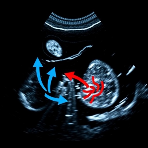
In a groundbreaking advancement in hepatocellular carcinoma (HCC) diagnostics, researchers have unveiled the significant potential of high frame rate contrast-enhanced ultrasound (H-CEUS) as a predictive tool for tumor biology and patient outcomes. This innovative imaging technique transcends the conventional ultrasound technology by delivering rapid, real-time visualization of tumor perfusion dynamics, providing unprecedented insights into the vascular characteristics intricately linked to tumor aggressiveness and recurrence risk.
Hepatocellular carcinoma, a primary malignancy of the liver, remains a formidable global health challenge due to its often-late diagnosis and high recurrence rates post-surgery. The conventional imaging modalities have struggled to offer detailed biological characterization preoperatively, impeding personalized treatment and effective prognostication. The introduction of H-CEUS addresses this gap by capturing subtle vascular patterns that correlate with tumor pathology at a molecular level, heralding a new era of non-invasive precision oncology.
This study, conducted over a three-year period from April 2021 to April 2024, involved 105 patients with pathologically confirmed HCC slated for radical surgical resection. Utilization of H-CEUS prior to surgery allowed for a meticulous assessment of microvascular architecture and perfusion kinetics, metrics that proved vital in distinguishing between tumor differentiation grades. The imaging uncovered distinct vascular morphologies, notably arborescent and fine vascular patterns, that were intimately associated with the biological behavior of the tumors.
The arborescent vascular morphology emerged as a critical imaging biomarker, independently predicting the presence of microvascular invasion (MVI), elevated proliferation indices indicated by high Ki-67 levels, and positive glypican-3 (GPC-3) expression. These factors have long been established as hallmarks of aggressive tumor biology and poor prognosis in HCC. By contrast, the fine vascular morphology correlated with more favorable characteristics, including absence of MVI, lower Ki-67 levels, and lack of GPC-3 expression, suggesting a less invasive tumor phenotype.
Further refinement of diagnostic granularity was achieved through the Liver Imaging Reporting and Data System (LI-RADS), where poorly differentiated tumors and those exhibiting MVI frequently corresponded to the LR-M category, which encompasses observations suspicious for malignancy. The study also elucidated differences in the feeding artery’s appearance concerning GPC-3 status, marking another layer of complexity in interpreting tumor vascular supply and its relationship with tumor biology.
The prognostic implications of these imaging findings were profound. Over a median follow-up duration of 17 months, patients exhibiting the arborescent vascular pattern demonstrated significantly shorter recurrence-free survival compared to those with fine vascular patterns. This highlights H-CEUS’s capacity not only to enhance diagnostic precision but also to provide crucial prognostic information that can influence clinical decision-making and postoperative surveillance strategies.
Interestingly, despite the diagnostic value of LI-RADS categories and feeding artery characteristics, these parameters did not independently predict recurrence-free survival, reinforcing the unique predictive strength of vascular morphology captured by H-CEUS. This insight challenges existing paradigms and suggests that dynamic vascular imaging may be a superior predictor of tumor biology and patient outcomes.
Of notable importance, when H-CEUS-derived vascular morphology data were combined with serum alpha-fetoprotein (AFP) levels—a well-known but imperfect biomarker for HCC—the predictive accuracy for tumor recurrence markedly improved. This synergistic approach yielded an area under the receiver operating characteristic curve (AUC) of 0.813, signaling robust diagnostic performance and endorsing a multimodal evaluation framework in clinical practice.
The clinical ramifications of these findings are expansive. Early and accurate identification of high-risk HCC patients enables tailored therapeutic strategies including more aggressive surgical approaches, adjuvant therapies, or intensified postoperative monitoring to mitigate recurrence risks. Moreover, H-CEUS is a radiation-free, cost-effective, and widely accessible modality, which potentiates its implementation across diverse healthcare settings, including resource-constrained environments.
Technically, the high frame rate involves capturing ultrasound images at exceptionally rapid intervals, thereby resolving the temporal resolution barriers that have limited previous contrast-enhanced ultrasound applications. This capability facilitates detailed real-time tracking of contrast agents as they traverse tumor microcirculation, revealing intricate vascular patterns that are invisible to standard imaging techniques.
The vascular morphology evaluation hinges on sophisticated image analysis discerning the branching complexity, density, and distribution of microvessels within the tumor matrix. Arborescent patterns reflect a tangled, irregular vasculature often associated with neoangiogenesis—a hallmark of malignant progression—while fine vascular patterns denote sparse and orderly vessel architecture, indicative of less aggressive pathology.
This study also highlights the intersection of imaging with molecular oncology, as the imaging phenotypes correlate with molecular markers such as Ki-67, a marker of cellular proliferation, and GPC-3, a membrane-bound proteoglycan implicated in HCC oncogenesis. This biomedical cross-talk enhances the understanding of tumor heterogeneity and offers a non-invasive window into tumor biology.
The sustained follow-up and rigorous pathological correlation in this research provide compelling evidence for integrating H-CEUS into the preoperative evaluation algorithm for HCC. The application of this modality could revolutionize oncologic imaging by shifting paradigms from purely anatomical assessments to functional and biological characterizations that directly inform prognosis and therapy.
In an oncology landscape increasingly favoring precision and personalization, H-CEUS stands as a beacon of innovation, merging cutting-edge imaging physics with clinical oncology to improve outcomes for patients battling hepatocellular carcinoma. Future directions may involve the integration of artificial intelligence algorithms to automate vascular morphology analysis and enhance predictive accuracy further.
This advancement invites a re-examination of clinical protocols and fosters collaborative efforts between radiologists, oncologists, and surgeons to harness the full potential of H-CEUS. The translation from bench to bedside promises earlier interventions, better survival chances, and personalized care trajectories for a patient population in dire need of improved therapeutic avenues.
Ultimately, this research underscores the critical importance of dynamic imaging modalities in cancer diagnostics, inaugurating a new chapter where visualizing the invisible—tumor biology at the microvascular level—equips clinicians with actionable intelligence to outmaneuver hepatocellular carcinoma recurrence and progression.
Subject of Research: High frame rate contrast-enhanced ultrasound for biological characterization and outcome prediction in hepatocellular carcinoma.
Article Title: High frame rate contrast-enhanced ultrasound in hepatocellular carcinoma: biological characteristics and patient outcomes.
Article References: Zhu, L., Li, N., Liang, S. et al. High frame rate contrast-enhanced ultrasound in hepatocellular carcinoma: biological characteristics and patient outcomes. BMC Cancer 25, 1488 (2025). https://doi.org/10.1186/s12885-025-14907-1
Image Credits: Scienmag.com
DOI: https://doi.org/10.1186/s12885-025-14907-1


