


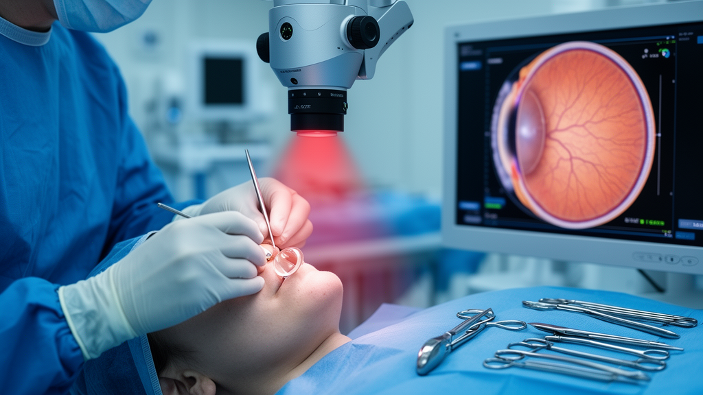
For the millions of people considering surgical correction of moderate to high myopia, the implantable Collamer lens, or ICL, has become one of the most talked-about options in modern ophthalmology. Unlike laser procedures that permanently reshape the cornea, ICL surgery places a foldable phakic lens inside the eye, behind the iris and in front of the natural crystalline lens. It preserves corneal tissue, is technically reversible, and delivers excellent visual outcomes, with high satisfaction reported across long-term follow-up studies. Yet like any intraocular operation, it is not risk-free. A new study from Wuhan Bright Eye Hospital in China has now revealed, with unprecedented real-time detail, a hidden mechanical injury occurring at the corneal incision during nearly half of all ICL procedures — an injury so small that conventional surgical microscopes cannot see it.
The injury in question is Descemet membrane detachment, or DMD. The Descemet membrane is a thin, resilient basement membrane that sits between the posterior corneal stroma and the corneal endothelium, the innermost cellular layer of the cornea. During intraocular surgery, mechanical forces can cause this membrane to peel away from the overlying stroma, creating a potential space that may fill with aqueous humor or viscoelastic material. Small, peripheral detachments often resolve spontaneously without any visual consequence. But larger or centrally located detachments can produce persistent corneal edema, delayed visual recovery, and, in severe cases, permanent corneal decompensation requiring surgical reattachment or even endothelial keratoplasty. In cataract surgery, clinically significant DMD has been reported in roughly 0.5 percent of phacoemulsification cases and up to 2.5 percent of extracapsular extractions. What has remained unclear until now is how often, and at what surgical moment, DMD develops during ICL implantation — a procedure that, on the surface, involves no ultrasonic energy and far fewer surgical steps.
To answer that question, the research team turned to a technology that is transforming how surgeons see inside the eye: intraoperative optical coherence tomography, or iOCT. Conventional operating microscopes illuminate only the corneal surface, making tiny, peripheral, or localized detachments nearly impossible to identify, especially once mild corneal edema sets in. Anterior segment OCT performed after surgery has become the gold standard for diagnosing DMD, but it captures only a static snapshot of what has already happened. The iOCT navigation microscope used in this study — the RESCAN 700 system from Carl Zeiss — integrates spectral-domain OCT directly into the surgical microscope, sweeping an OCT crosshair systematically from one end of the incision to the other while the operation proceeds. This allowed the surgical team to observe, in real time and at every step, whether the Descemet membrane was separating at the incision site and whether the extent of any detachment was changing as the procedure progressed.
The study was a single-center retrospective case series involving 100 eyes of 50 patients, aged 21 to 38 years, all of whom underwent ICL surgery between July and October 2024. Every procedure was performed by a single experienced surgeon using a 3.0-millimeter steep-axis clear corneal incision. The surgical workflow comprised four distinct steps: creation of the corneal tunnel incision with a disposable keratome; insertion of the foldable ICL through the incision using a loaded injector cartridge, with injection of a sodium hyaluronate viscoelastic agent to maintain anterior chamber depth; positioning of the lens haptics into the ciliary sulcus behind the iris using an adjustment hook; and finally removal of the viscoelastic agent via irrigation and aspiration. At each step, the iOCT system recorded whether DMD had occurred at the incision and whether previously detected detachments had extended. All procedures were completed successfully without intraoperative complications.
The findings were striking. No DMD was detected by the end of incision creation, indicating that the initial cut itself, when performed cleanly, does not disturb the Descemet membrane. But as the operation progressed, the cumulative proportion of eyes with DMD climbed steadily. By the end of ICL insertion, DMD had been detected in 21 percent of eyes. By the end of lens positioning, the cumulative figure had risen to 34 percent. And by the end of viscoelastic agent removal, 45 percent of eyes had developed DMD at some point during the procedure. The pattern of involvement shifted across steps as well. During ICL insertion, detachments overwhelmingly involved the anterior lip of the incision — 18 of 21 eyes — consistent with friction between the delivery device and the front edge of the corneal tunnel. During lens positioning, the pattern reversed, with 32 of 34 eyes showing posterior lip involvement, suggesting contact between the positioning hook and the back edge of the incision. Only two eyes developed detachments involving both lips simultaneously, both during viscoelastic removal, possibly related to irritation of the tunnel by the irrigation needle and pressure fluctuations caused by fluid exchange in the anterior chamber.
To identify risk factors, the researchers employed a sophisticated statistical approach. Because both eyes from the same patient were included, eye-level observations could not be treated as independent; a mixed-effects model with a patient-specific random intercept was used to account for the correlation between fellow eyes. A two-part model was constructed to handle the substantial zero inflation in the DMD extent data: the first part analyzed the odds of DMD occurrence using multilevel logistic regression, while the second part, conditional on the presence of DMD, analyzed factors influencing its severity using multilevel linear regression on log-transformed values. Variable selection was performed using LASSO regression, a method suited to small samples and high-dimensional data. The analysis revealed one statistically significant predictor: lower preoperative intraocular pressure was associated with higher odds of DMD occurrence, with an odds ratio of 0.77 per 1-millimeter-of-mercury increase in pressure (95 percent confidence interval, 0.61 to 0.97; P = 0.03). In other words, eyes with softer, lower-pressure corneas were more vulnerable to mechanical detachment at the incision. Notably, none of the evaluated factors — including age, cylindrical power, axial length, and anterior chamber depth — was significantly associated with the extent of detachment once it occurred.
The clinical implications of these findings are considerable. Because DMD was most likely to develop during ICL insertion and during viscoelastic removal, the study points to specific, actionable strategies for reducing risk. Surgeons encountering significant resistance while creating the incision should immediately switch to a sharper blade rather than force the current one through. The incision size should match the injector cartridge as closely as possible, because an excessively tight incision increases friction between the injector and the corneal tunnel. The volume of viscoelastic agent injected should be minimized, since thorough removal of a high-molecular-weight sodium hyaluronate gel was associated with the largest number of detachments and the most prolonged cases. For patients with lower preoperative intraocular pressure, particular attention should be paid to maintaining a relatively high intraoperative pressure, which may reduce the mechanical vulnerability of the Descemet membrane. Throughout the procedure, careful instrument manipulation and minimization of repeated contact with the corneal incision remain the most fundamental preventive principles.
It is important to contextualize the incidence figures reported here. The overall proportion of eyes with DMD at some point during ICL surgery was lower than the 96 percent incidence observed in cataract surgery at Zhongshan Ophthalmic Center using similar real-time imaging, a discrepancy the authors attribute to the smaller number of surgical steps in phakic lens implantation and the absence of ultrasonic energy. Moreover, the high figures in both ICL and cataract series reflect the sensitivity of iOCT, which detects small, clinically silent detachments that would be missed on routine slit-lamp examination. In conventional clinical practice, where DMD is diagnosed postoperatively, reported incidences are far lower — often below 0.5 percent — precisely because subclinical detachments go unrecognized. The real-time approach used in this study therefore reveals a hidden burden of injury that has always been present but never before visible during the operation itself.
The study has limitations that temper its conclusions. All procedures were performed by a single experienced, high-volume surgeon, which ensures technical standardization but may have contributed to a relatively low DMD incidence and restricts generalizability to less experienced operators. A recent study comparing phacoemulsification performed by residents versus professional staff reported a higher incidence of incision-site DMD in the resident group, supporting surgeon experience as an important determinant of this complication. All DMD measurements were obtained using ImageJ software and evaluated by a single observer, introducing potential observer-related variability. Patients with poor corneal or ocular surface conditions were excluded, limiting applicability to compromised corneas. The single-center design, relatively small sample size, and limited follow-up duration further constrain the precision of the estimates. Future multicenter prospective studies incorporating larger samples, surgeons of varying experience levels, standardized assessment by multiple masked observers, and longer follow-up are needed to confirm and extend these findings.
Nevertheless, the study represents a milestone in the application of real-time imaging to refractive surgery. To the authors’ knowledge, it is the first investigation to use intraoperative OCT navigation to dynamically assess changes in DMD at a 3.0-millimeter surgical incision during phakic intraocular lens implantation. By revealing exactly when, where, and why the Descemet membrane separates during ICL surgery, the work transforms an invisible complication into a visible, measurable, and preventable event. For the growing population of patients choosing ICL implantation as an alternative to laser vision correction, and for the surgeons who operate on them, these findings offer a clear message: the corneal incision is not merely a doorway through which instruments pass, but a delicate structure whose integrity must be actively protected at every step of the procedure.
Subject of Research: Ophthalmology
Article Title: Intraoperative OCT observation of incision-related descemet membrane detachment during implantable collamer lens implantation
Article References: Xie, J., Guan, N., Liu, Y., Zheng, Z., He, X., & Shen, Z. (2026). Intraoperative OCT observation of incision-related descemet membrane detachment during implantable collamer lens implantation. Frontiers in Medicine, 13, Article 1888213. https://doi.org/10.3389/fmed.2026.1888213
Image Credits: AI Generated
DOI: 10.3389/fmed.2026.1888213
Keywords: implantable Collamer lens, Descemet membrane detachment, intraoperative OCT, refractive surgery, myopia, corneal incision, viscoelastic agent, intraocular pressure, surgical imaging, ophthalmology, Intraoperative, observation
Cite Scienmag News
Ophelia Keating. (October 15, 2026). Hidden corneal injury during lens implantation caught in real time by surgical OCT. Scienmag. https://scienmag.com/hidden-corneal-injury-during-lens-implantation-caught-in-real-time-by-surgical-oct/
Ophelia Keating. "Hidden corneal injury during lens implantation caught in real time by surgical OCT." Scienmag, 15 October 2026, https://scienmag.com/hidden-corneal-injury-during-lens-implantation-caught-in-real-time-by-surgical-oct/. Accessed 15 October 2026.
Ophelia Keating. "Hidden corneal injury during lens implantation caught in real time by surgical OCT." Scienmag. October 15, 2026. https://scienmag.com/hidden-corneal-injury-during-lens-implantation-caught-in-real-time-by-surgical-oct/


