


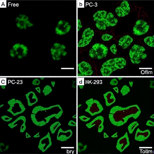
A common plant-derived molecule has gained a high-tech delivery system—and, in laboratory experiments, the combination appeared to suppress prostate cancer cells more effectively over time than the unencapsulated compound. Researchers at the University of Mazandaran in Iran tested gallic acid, a polyphenol found in many foods and medicinal plants, after loading it into biodegradable chitosan–poly(acrylic acid) nanoparticles. The nanoformulation reduced the metabolic activity of PC-3 prostate cancer cells and altered the expression of genes associated with cell death and cell-cycle control. The findings, published in 3 Biotech, suggest that a carefully engineered carrier could change how a natural compound reaches and affects cancer cells. They do not yet demonstrate a treatment for patients: the work was performed in cultured cells, and the biological behavior of the particles inside a living body remains unknown.
Gallic acid is a small phenolic molecule with antioxidant and other biological activities, but its potential as an anticancer agent is limited by the familiar problems facing many natural compounds. A molecule can show promising effects in a dish yet fail to reach the right tissue, remain stable long enough, or enter cells at an effective concentration. Nanoparticles are designed to address some of those barriers by packaging an active substance inside a protective structure that can influence its solubility, stability, cellular uptake and release. In this study, Fatemeh Fathali, Ali Taravati and Majid Tafrihi used chitosan, a positively charged polymer derived from chitin, together with poly(acrylic acid), a negatively charged polymer. Their opposite electrical charges allow the polymers to assemble through electrostatic attraction into a polyelectrolyte complex capable of holding gallic acid.
The optimized particles had a mean hydrodynamic diameter of 310 ± 33 nanometres, a surface charge, or zeta potential, of +8.8 millivolts, and an encapsulation efficiency of 67 percent. Hydrodynamic diameter is the effective size of a particle as it moves through liquid, including the layer of solvent and ions that travel with it; it is typically measured using dynamic light scattering. The value is therefore not necessarily identical to the dry particle size seen under an electron microscope. Zeta potential provides an estimate of the electrical potential near the particle surface and can help indicate how colloids interact with one another in suspension. A modestly positive charge may influence interactions with negatively charged cell membranes, although surface charge alone does not predict how a nanoparticle will behave in blood or tissue. The 67 percent encapsulation efficiency means that roughly two-thirds of the gallic acid used during formulation became associated with the nanoparticle system under the reported conditions.
The carrier was also designed to release its cargo gradually rather than releasing all of the gallic acid at once. Release experiments showed sustained, pH-dependent behavior under different physiological conditions. That feature arises from the chemistry of the polymer network. Chitosan contains amino groups whose charge changes with acidity, while poly(acrylic acid) contains carboxyl groups that can become more or less ionized as pH changes. Because ionization affects the strength of the electrostatic interactions between the two polymers, it can alter swelling, compactness and the diffusion of gallic acid out of the particles. In principle, such responsiveness could allow the formulation to behave differently in environments with different acidity. In practice, however, release profiles measured in laboratory solutions are only an early approximation of what happens amid proteins, enzymes, immune cells and changing fluid flows inside the body.
To test biological activity, the researchers exposed PC-3 cells to free gallic acid and to gallic acid carried by the chitosan–poly(acrylic acid) nanoparticles. PC-3 is a widely used human prostate cancer cell line, originally established from a metastatic prostate tumor and commonly employed to study cancer-cell survival, migration and drug response. The team also examined HEK-293 cells, a human embryonic kidney-derived line, as a comparison model for cells that were not prostate cancer cells. Cell viability was estimated with the MTT assay, in which metabolically active cells convert a yellow tetrazolium compound into purple formazan crystals. The amount of colored product is used as an indirect measure of metabolic activity. It is important to note that MTT does not count living cells directly and does not identify a specific death mechanism; changes in metabolism can influence the signal even when cell number and viability are not changing in exactly the same way.
The strongest evidence for a time-dependent effect came from the half-maximal inhibitory concentration, or IC50—the concentration required under a specified experimental condition to reduce the measured response by 50 percent. After 24 hours, the IC50 for the nano-encapsulated gallic acid was 97 micrograms per millilitre, compared with 44 micrograms per millilitre for free gallic acid. At that early time point, free gallic acid therefore produced the stronger apparent effect in the MTT readout. After 48 hours, however, the IC50 for the nanoparticle formulation fell to 37 micrograms per millilitre, while the value for free gallic acid was 41 micrograms per millilitre. The shift suggests that encapsulation may have slowed the initial availability of the compound while sustaining its biological influence over a longer period. It is not evidence that the nanoparticles are universally more potent, and the numerical difference at 48 hours should be interpreted within the variability and experimental design of the cell assay.
HEK-293 cells showed lower sensitivity than PC-3 cells under the tested conditions, a result that may indicate some degree of differential response between the cancer model and the comparison line. Yet HEK-293 cells are not a normal prostate-cell model, so the comparison cannot establish cancer selectivity or safety for healthy prostate tissue. Cancer and noncancerous cells can differ in metabolism, membrane composition, stress responses and drug transport, but cell-line comparisons are an imperfect substitute for testing primary human cells or tissue models. Nanoparticles can also interfere with colorimetric assays by absorbing light, scattering it, or interacting with assay reagents. Consequently, the MTT findings are most useful as an initial signal that should be confirmed with independent measurements, such as direct cell counting, membrane-integrity assays, clonogenic survival tests and analyses that distinguish apoptosis from necrosis.
Additional experiments pointed toward changes in pathways controlling programmed cell death and proliferation. Treated PC-3 cells showed increased expression of TP53 and BAX, alongside reduced expression of BCL2 and CCND1. TP53 encodes p53, a stress-responsive protein that can pause the cell cycle or promote programmed cell death when a cell experiences severe DNA damage or other threats. BAX is a pro-apoptotic member of the BCL2 protein family and can help promote mitochondrial outer-membrane permeabilization, an important step in intrinsic apoptosis. BCL2 generally supports cell survival by opposing that mitochondrial death pathway. CCND1 encodes cyclin D1, a regulator that helps drive progression through the early stages of the cell cycle. The expression pattern is therefore compatible with increased apoptotic signaling and reduced proliferative drive. But gene-expression changes alone do not prove that apoptosis occurred. Confirmation would require measurements of proteins, caspase activation, mitochondrial changes, DNA fragmentation or other functional markers of cell death.
The researchers also used a wound-healing assay to examine how treatment affected the ability of cells to close an artificial gap in a cell layer. In this test, a scratch is made across a confluent population and the narrowing of the gap is tracked over time. A slower rate of closure can reflect reduced cell migration, reduced cell proliferation, or both. The treated PC-3 cultures showed reduced wound closure, but the study appropriately cautions that the result cannot be assigned solely to an antimetastatic effect. If a treatment lowers cell metabolism or viability, fewer cells remain available to divide and move into the gap. More specialized migration assays, including methods that block proliferation or use defined barriers, would be needed to separate these processes. The finding nevertheless adds a second dimension to the viability results by indicating that the formulation affected a behavior relevant to tumor progression in vitro.
The appeal of the system lies in the way formulation and biology intersect. Gallic acid supplies the active chemical signal, while the chitosan–poly(acrylic acid) matrix changes when and how that signal becomes available. A sustained-release carrier could potentially reduce rapid loss of a soluble compound and maintain exposure near cells, but a nanoparticle that works in a dish must still pass a far higher bar before it can be considered a medical technology. Its stability in physiological fluids, interaction with serum proteins, distribution through the body, uptake by tumors, degradation products, immune effects and clearance would all need to be established. Particle size and surface charge can influence uptake by cells and removal by organs, while the tumor microenvironment is heterogeneous and does not reliably guarantee nanoparticle accumulation. The study did not test animals, tumors, clinical samples or combination treatment with existing prostate-cancer drugs.
For now, the results are best understood as a formulation study with a promising laboratory signal rather than a ready-made natural cancer therapy. The nanoparticle preparation achieved measurable gallic-acid loading, pH-responsive release and delayed activity against PC-3 cells, while the associated gene-expression changes offered clues about possible mechanisms. The work also highlights why claims about “nanotechnology cures” require restraint: a lower IC50 in cultured cells does not automatically translate into improved survival, selective tumor targeting or safety in people. The next steps would include independent validation with multiple prostate-cancer and noncancerous cell models, mechanistic assays confirming apoptosis, rigorous controls for nanoparticle interference, pharmacokinetic studies and animal testing. Only after those stages could researchers determine whether these biodegradable polymer particles can turn gallic acid’s biochemical potential into a practical component of prostate-cancer treatment.
Cite Scienmag News
Juliet Wilcox. (August 28, 2026). Free versus nano-encapsulated gallic acid compared in PC-3 and HEK-293 cells. Scienmag. https://scienmag.com/free-versus-nano-encapsulated-gallic-acid-compared-in-pc-3-and-hek-293-cells/
Juliet Wilcox. "Free versus nano-encapsulated gallic acid compared in PC-3 and HEK-293 cells." Scienmag, 28 August 2026, https://scienmag.com/free-versus-nano-encapsulated-gallic-acid-compared-in-pc-3-and-hek-293-cells/. Accessed 3 September 2026.
Juliet Wilcox. "Free versus nano-encapsulated gallic acid compared in PC-3 and HEK-293 cells." Scienmag. August 28, 2026. https://scienmag.com/free-versus-nano-encapsulated-gallic-acid-compared-in-pc-3-and-hek-293-cells/


