


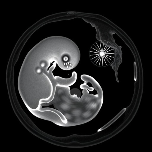
In a groundbreaking advancement poised to transform forensic and pathological investigations, researchers have unveiled their initial experiences utilizing a 0.31 Tesla low-field magnetic resonance imaging (MRI) system in post-mortem examinations of fetuses. This pioneering study, conducted by Gascho, Kuntze, Deininger-Czermak, and colleagues, marks a significant milestone in prenatal forensic diagnostics, offering a non-invasive, detailed imaging alternative when traditional autopsies are constrained or declined. The low-field MRI approach introduces new dimensions of accessibility and practicality in post-mortem fetal imaging, setting the stage for a paradigm shift in both clinical and legal medicine.
The study addresses a longstanding challenge in perinatal and forensic pathology: how to achieve high-resolution imaging that can reveal anatomical and pathological details in fetal tissues without resorting to invasive autopsy procedures. Typically, high-field MRI units, operating at 1.5 Tesla or above, are employed for advanced diagnostic imaging, but these systems are expensive, bulky, and not widely available in many clinical or forensic settings. The use of a 0.31 Tesla low-field MRI unit emerges as a cost-effective, portable, and efficient alternative, broadening the scope of fetal post-mortem examinations.
At the core of this innovative research lies the technical capability to capture detailed morphological information from deceased fetal subjects with minimal artifact interference, despite the lower magnetic field strength. Lower fields generally present challenges in signal-to-noise ratio (SNR) and spatial resolution, but the researchers ingeniously optimized imaging protocols to counterbalance these limitations. Their work demonstrated that carefully tuned sequences and advanced image processing techniques can substantially enhance tissue contrast and fine structural visibility at 0.31 Tesla.
One of the notable technical achievements highlighted in the research was the optimization of T1-weighted and T2-weighted imaging sequences tailored specifically for fetal tissues post-mortem. These sequences were calibrated to exploit the relaxation properties unique to fetal anatomy and developmental stages, thereby maximizing contrast differentiation between critical structures such as brain tissue, thoracic organs, and musculoskeletal elements. This methodological refinement is crucial because post-mortem tissue characteristics differ markedly from those in living subjects, often complicating conventional MRI interpretations.
Furthermore, the study underscores the portability and reduced operational demands of low-field MRI systems, which enable broader implementation in forensic pathology settings beyond large urban centers and academic hospitals. Unlike high-field MRI machines, low-field units require less shielding, consume less power, and produce less acoustic noise, thereby facilitating easier integration into morgues and forensic laboratories. This accessibility could revolutionize how fetal deaths are investigated, particularly in resource-limited or geographically isolated regions.
Beyond physical logistics, the ethical implications of using non-invasive imaging in fetal post-mortem exams are profound. Many families decline conventional autopsies for cultural, religious, or personal reasons, often leaving medical professionals without definitive answers regarding causes of fetal demise. Employing low-field MRI as a minimally invasive modality respects these sensitivities while generating diagnostic insights that may inform parental counseling, epidemiological tracking, and medico-legal investigations.
The authors further observed that the image quality, while naturally not equivalent to that of high-field systems, was sufficiently robust to identify major congenital anomalies, brain malformations, and thoracoabdominal abnormalities. In several instances, the low-field MRI findings correlated strongly with clinical histories and available biochemical data, suggesting a promising diagnostic concordance that warrants further validation in larger cohorts.
Importantly, this research opens pathways to more extensive studies that could calibrate low-field MRI to detect subtler pathologies such as microstructural brain injuries, placental abnormalities, or signs of intrauterine infections. The adaptability of the imaging protocols and the potential enhancement through emerging AI-assisted image reconstruction methods could amplify the resolution and interpretative accuracy of post-mortem imaging in near future applications.
The authors also discuss how the particular magnetic environment of 0.31 Tesla might reduce certain imaging artifacts commonly encountered in high-field MRI, such as susceptibility effects near air-tissue interfaces. This factor may enhance visualization of delicate fetal structures, potentially overcoming some technical barriers that have historically limited fetal MRI.
In addition to post-mortem diagnostics, the low-field MRI technology described in this study bears implications for clinical prenatal imaging, especially in high-risk pregnancies where routine MRI is constrained by accessibility or safety concerns. The development trajectory of compact, low-field MRI scanners could ultimately enrich prenatal care by enabling bedside imaging or even ambulatory scan capabilities in the near future.
Collaboration between forensic pathologists, radiologists, and MRI physicists was pivotal for the success of this initiative, as it required an intricate understanding of fetal pathology combined with technical expertise in MRI physics to customize the imaging sequences and interpret acquired data within clinically meaningful frameworks. This interdisciplinary approach exemplifies how innovation at the interface of technology and medicine can lead to impactful solutions to complex healthcare challenges.
While the study acknowledges limitations relating to the relatively small sample size and the need for further comparative studies involving both high- and low-field MRI data, the preliminary findings already suggest a valuable role for low-field systems in forensic medicine workflows. Particular emphasis is placed on the potential to reduce autopsy rates, improve family acceptance, and expedite forensic investigations, which in turn could alleviate systemic burdens in medicolegal death investigations.
Moreover, this technological advance carries significant ramifications for global health. With many regions lacking access to high-field MRI infrastructure, the democratization of post-mortem fetal imaging through affordable low-field units could help bridge gaps in neonatal mortality surveillance and research, ultimately informing public health policies aimed at reducing stillbirth rates and improving maternal-fetal care.
The ingenuity of employing low-field MRI technology at 0.31 Tesla in this context offers a compelling example of how reimagined uses of existing technologies can address unmet medical dilemmas. By enhancing post-mortem fetal examination capabilities, this approach serves not only scientific inquiry but also ethical considerations and practical constraints, all while expanding diagnostic reach in a traditionally underserved space.
As this initially published research gains traction, it is expected to inspire further investigation into optimizing low-field MRI protocols, integrating machine learning for image enhancement, and exploring combined multimodal imaging techniques. These advancements may well redefine the standards of fetal post-mortem diagnostics and set new benchmarks for forensic methodology in the years to come.
In summary, the first experiences reported with 0.31 Tesla low-field MRI in post-mortem fetal examinations have unveiled a promising frontier where technical innovation intersects with compassionate medical practice. This study offers a glimpse into a future where detailed fetal assessments become more accessible, respectful, and accurate, significantly impacting the fields of forensic pathology, legal medicine, and prenatal healthcare at large.
Cite Scienmag News
Ophelia Keating. (December 26, 2025). First Use of 0.31T MRI in Fetal Autopsy. Scienmag. https://scienmag.com/first-use-of-0-31t-mri-in-fetal-autopsy/
Ophelia Keating. "First Use of 0.31T MRI in Fetal Autopsy." Scienmag, 26 December 2025, https://scienmag.com/first-use-of-0-31t-mri-in-fetal-autopsy/. Accessed 6 September 2026.
Ophelia Keating. "First Use of 0.31T MRI in Fetal Autopsy." Scienmag. December 26, 2025. https://scienmag.com/first-use-of-0-31t-mri-in-fetal-autopsy/


