


…
…
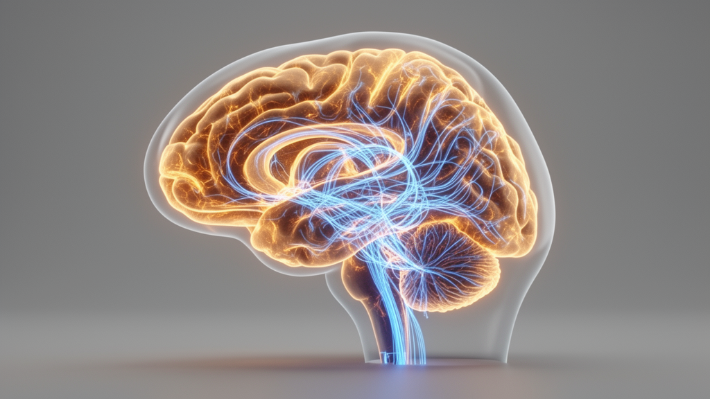
The shift from viewing dystonia as a localized circuit defect to understanding it as a distributed network disorder has profound implications for how clinicians and researchers interpret the growing body of imaging and electrophysiological data. When Bhatia and colleagues surveyed the lesion literature and found that dystonia could arise from damage to heterogeneous brain regions, the observation only made coherent sense once those regions were understood as nodes within shared large-scale circuits. A lesion in the putamen, a lesion in the thalamus, and a lesion in the cerebellum can each produce overlapping clinical phenomenology not because the structures are functionally interchangeable in isolation, but because each participates in the same recurrent loops that ultimately shape the output of the motor system. This network logic also explains why routine clinical scans are so frequently unrevealing in dystonia patients. The pathology in many forms of the disorder is not a gross structural lesion but a subtle derangement of how regions communicate with one another, a derangement that structural computed tomography and conventional magnetic resonance imaging sequences were never designed to detect.
The distinction between structural and functional abnormality deserves particular emphasis because it shapes diagnostic expectations. Voxel-based morphometry studies, which quantify gray matter volume across the whole brain, have reported modest volume differences in the sensorimotor cortex, basal ganglia, cerebellum, and thalamus in various dystonia cohorts, but these differences are typically small and inconsistent across samples. Diffusion imaging, which probes the integrity of white matter pathways by measuring the directional diffusion of water molecules, has similarly revealed subtle alterations in tracts connecting the nodes of the motor networks. The very subtlety of these findings reinforces the conclusion that dystonia is primarily a disorder of network dynamics rather than of network architecture. A brain can possess structurally intact connections yet still route information through those connections abnormally, much as a road network can be physically sound while traffic flows along it in a disorganized pattern. Functional imaging modalities capture this dynamic dimension directly, and the convergence of PET, functional MRI, EEG, magnetoencephalography, and functional near-infrared spectroscopy evidence on the same cortico-striato-pallido-thalamocortical and cerebello-thalamocortical loops provides a degree of multimodal consistency that no single modality could achieve alone.
PET studies occupy a distinctive position within this multimodal landscape because they can measure not only metabolic activity but also neurotransmitter systems in vivo. The ability to image dopamine, GABA, and acetylcholine signaling directly connects the network-level abnormalities observed in dystonia with the molecular-level pathophysiology suggested by genetic and pharmacological evidence. Dopamine receptor abnormalities, reduced GABAergic inhibition, and cholinergic dysfunction have each been implicated in dystonia through different lines of investigation, and PET provides the means to observe these systems operating within the living human brain. This molecular-to-network bridge is essential because it prevents the network perspective from becoming purely descriptive. Knowing that a particular loop is dysfunctional is one thing; knowing which neurotransmitter system within that loop is deranged is quite another, and the latter knowledge opens the door to pharmacological interventions that could complement neuromodulation approaches.
The invasive recordings obtained during deep brain stimulation surgery represent a qualitatively different level of measurement precision compared to non-invasive imaging. While functional MRI offers millimeter-scale spatial resolution, its temporal resolution is limited by the hemodynamic response, which unfolds over seconds. EEG and magnetoencephalography offer millisecond temporal resolution but with limited spatial precision. Recordings from DBS electrodes, by contrast, combine high temporal resolution with direct measurement of local field potentials from deep brain structures that are otherwise inaccessible in humans. These recordings have revealed pathological oscillatory activity in the beta and gamma frequency ranges within the basal ganglia and thalamus of dystonia patients, and analyses of stimulus-evoked responses have begun to map effective connectivity, meaning the directed influence of one region on another, within the affected circuits. The concept of effective connectivity is particularly important because it moves beyond correlation. Resting-state functional connectivity can tell us that two regions are co-active, but effective connectivity tells us whether activity in one region drives activity in the other, and in which direction. This directional information is what a network model of pathophysiology ultimately requires.
Non-invasive brain stimulation techniques offer a complementary approach to network interrogation that is fundamentally perturbative rather than observational. Transcranial magnetic stimulation applies brief magnetic pulses that induce electrical currents in the underlying cortex, transiently disrupting or modulating local activity. Transcranial electrical stimulation uses weak currents applied through scalp electrodes to achieve similar effects. Transcranial ultrasound stimulation, a more recent addition to the toolkit, uses focused ultrasound waves to modulate neural activity with potentially greater spatial precision than magnetic or electrical methods. The perturbative logic of these techniques is powerful: if a brain region is a critical node in a dysfunctional network, then transiently disrupting that region should produce measurable changes in network behavior, and those changes can reveal the region’s causal role. Beyond their diagnostic utility, these stimulation methods also carry therapeutic potential. Applications designed to decrease cortical excitation, increase inhibition, and normalize abnormal plasticity have shown promise in small studies, and the ability to combine stimulation with concurrent EEG recording enables closed-loop paradigms in which stimulation parameters are adjusted in real time based on the brain’s response.
The computational modeling platform known as The Virtual Brain represents a significant advance in the effort to integrate multimodal data into a coherent simulation of whole-brain dynamics. Rather than treating each imaging modality as a separate source of findings requiring separate interpretation, The Virtual Brain constructs a personalized model of an individual patient’s brain network using structural connectivity derived from diffusion imaging, then simulates the dynamic behavior that emerges from that architecture. The simulated activity can be compared against the patient’s actual functional imaging data, and discrepancies between simulation and observation can point to specific parameters of network dysfunction. This approach operates at the level of mean-field dynamics, a spatial abstraction that matches the resolution of most human imaging work, and it transforms the network perspective from a descriptive framework into a predictive one. A model that can reproduce a patient’s observed network abnormalities has the potential to predict how those abnormalities will respond to a given intervention, whether that intervention is a particular DBS target, a specific stimulation waveform, or a course of physiotherapy.
Across the clinical spectrum of dystonia, from focal forms affecting a single body region to generalized forms involving multiple body parts, and across the genetic etiologies that underlie many pediatric cases, the network perspective reveals both shared and distinct patterns of dysfunction. The shared patterns suggest common final pathways through which diverse etiologies converge on similar network-level derangements, which is consistent with the observation that symptomatic treatments such as DBS can be effective across multiple dystonia subtypes. The distinct patterns, meanwhile, suggest that phenotypic and genotypic subtypes may require different therapeutic targets or different stimulation parameters. This tension between convergence and divergence is a central theme in the network pathophysiology of dystonia, and resolving it will require the kind of multimodal, integrative approach that the review advocates.
Several understudied dimensions of dystonia network pathophysiology merit attention in future research. Longitudinal and developmental dynamics remain poorly characterized, particularly the question of how the dystonic network evolves over time in individual patients and how it differs between pediatric and adult-onset forms. The relationship between genotype and phenotype, as both axes become better characterized through objective measures, should provide a framework for understanding how specific genetic mutations give rise to specific network topographies. Motor behavior itself provides important constraints on circuit models, since overtrained movement patterns are thought to be a causal factor in many task-specific dystonias, and properly designed physiotherapy interventions should be able to modulate the network toward normalized function. Tremor in dystonia and functional dystonia represent two specific phenotypic aspects that warrant deeper investigation from a network perspective, as both may involve circuit-level mechanisms that overlap with, yet are distinct from, the core dystonic network pathology.
The translation of network insights into precision interventions is already underway. Circuit-guided neuromodulation, in which stimulation targets and parameters are selected based on an individual patient’s network profile, and closed-loop brain stimulation, in which stimulation is delivered only when pathological network states are detected, represent the leading edge of this translation. The convergence of improved imaging, higher-resolution invasive recordings, refined stimulation techniques, and increasingly sophisticated computational models creates the conditions for a genuinely personalized approach to dystonia treatment. Advancing these multimodal and integrative methodologies will be essential to unraveling the complex dynamics underlying dystonia and translating mechanistic insights into interventions that directly target the neural network basis of the disorder.
Subject of Research: Neuroscience
Article Title: Brain network pathophysiology in dystonia
Article References: Peterson, D. A., Kim, M., Chen, R., Eidelberg, D., Gallea, C., Horn, A. G., Lehericy, S., McIntosh, A. R., Perlmutter, J. S., Sadnicka, A., Sanger, T. D., Santarnecchi, E., Starr, P. A., Teller, J. K., Hallett, M., & Simonyan, K. (2026). Brain network pathophysiology in dystonia. Dystonia, 4, Article 15446. https://doi.org/10.3389/dyst.2025.15446
Image Credits: AI Generated
Keywords: dystonia, brain networks, deep brain stimulation, neuroimaging, neuromodulation, movement disorders, Brain, network, pathophysiology, scientific research
Cite Scienmag News
Cassandra Pierce. (October 16, 2026). Dystonia Rewired: How Brain Networks Explain a Movement Disorder. Scienmag. https://scienmag.com/dystonia-rewired-how-brain-networks-explain-a-movement-disorder/
Cassandra Pierce. "Dystonia Rewired: How Brain Networks Explain a Movement Disorder." Scienmag, 16 October 2026, https://scienmag.com/dystonia-rewired-how-brain-networks-explain-a-movement-disorder/. Accessed 16 October 2026.
Cassandra Pierce. "Dystonia Rewired: How Brain Networks Explain a Movement Disorder." Scienmag. October 16, 2026. https://scienmag.com/dystonia-rewired-how-brain-networks-explain-a-movement-disorder/


