


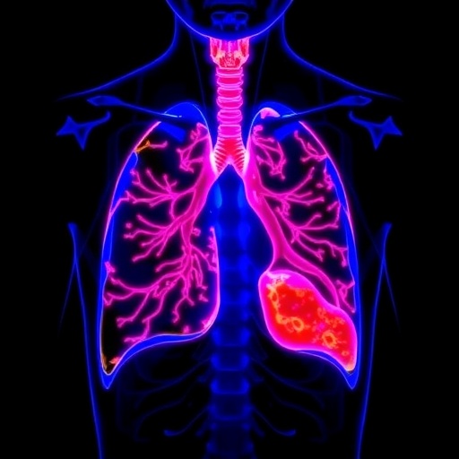
A new imaging platform designed to help surgeons see cancer more clearly during operations is bringing together two capabilities that have traditionally required separate cameras. The single-chip, dual-gain color–near-infrared system described by Zhu, Hajek, Jin and colleagues is engineered to capture ordinary visible-light information and fluorescence signals in the near-infrared region at the same time. Its intended applications are fluorescence-guided lung and breast cancer surgery, where the difference between healthy and malignant tissue can be subtle, the operating field can change rapidly, and even a small amount of residual disease may matter. The work, published in Light: Science & Applications, points toward a more compact form of surgical imaging that could place anatomical context and molecular contrast inside one synchronized view.
Fluorescence-guided surgery relies on a simple but powerful idea: a fluorescent compound is introduced into the body and accumulates preferentially in, or becomes activated around, a target such as a tumor. When illuminated with an appropriate wavelength, the compound emits light at a longer wavelength. In many surgical systems, that emission is detected in the near-infrared, where tissue can be relatively less absorbing than it is in visible wavelengths. The resulting signal can reveal structures that are difficult to distinguish by eye. Yet fluorescence alone is not always enough. A bright glowing region may not communicate its exact position relative to skin, blood vessels, instruments, or anatomical boundaries. A conventional color image supplies that context, but combining both views has often required multiple optical paths, cameras, or carefully aligned systems.
The camera introduced in this study addresses that problem at the sensor level. Rather than treating visible color and near-infrared fluorescence as entirely separate imaging tasks, it is designed as a single-chip platform capable of recording both. The “color” component refers to the wavelengths that generate familiar red, green, and blue information, allowing the system to reproduce the visual appearance of tissue and the surgical field. The “NIR” component extends sensitivity beyond the visible spectrum, enabling detection of fluorescence signals that human eyes cannot see. Because the two types of information are acquired by one integrated detector rather than by physically separate cameras, the architecture has the potential to reduce alignment errors, simplify the instrument, and maintain a more direct relationship between the fluorescence signal and the anatomy shown in the same frame.
The second defining feature is the camera’s dual-gain operation. Image sensors must cope with scenes that contain extremely different light levels. A surgical field may include dim fluorescence from a small target alongside intense reflected illumination from tissue, surgical lamps, or specular highlights. If a detector is configured for high sensitivity, bright regions can saturate; if it is configured for a broad range, weak signals may be buried in electronic noise. Dual-gain readout addresses this compromise by allowing the sensor to operate with two different signal-conversion conditions. A high-gain path can emphasize small numbers of collected photons and improve the visibility of weak fluorescence, while a lower-gain path can preserve information in brighter areas without reaching saturation as quickly. Combining these responses expands the usable dynamic range of the image.
That capability is particularly important in fluorescence-guided procedures because the clinically useful signal is not necessarily uniform. Tumor tissue may fluoresce strongly in one region and weakly at its edges, while the surrounding anatomy can reflect the excitation light at a much higher intensity. The camera therefore has to distinguish a meaningful molecular signal from background illumination, sensor noise, and optical leakage between channels. A dual-gain architecture does not by itself identify cancer, and it does not replace pathological analysis. Its role is to improve the quality and range of the visual information available to the surgical team. Better imaging can make a fluorescent boundary easier to interpret, but the decision about whether tissue is malignant still depends on the full clinical context and, ultimately, diagnostic evaluation.
Putting visible and near-infrared imaging on one chip also creates technical challenges. Silicon-based image sensors are naturally sensitive to visible light and can extend into part of the near-infrared, but the spectral responses of neighboring pixels or color channels can overlap. This overlap, known as spectral crosstalk, can contaminate color reproduction or make it harder to quantify fluorescence accurately. An imaging system intended for surgery must therefore be calibrated so that the intensity assigned to each channel reflects the underlying optical signal as faithfully as possible. Optical filters, pixel design, exposure control, signal processing, and correction algorithms can all influence the final image. The central engineering task is to retain recognizable color information while extracting a weak NIR signal without allowing the two measurement modes to interfere with one another.
The single-chip format could also matter in the operating room for reasons beyond image quality. Multi-camera systems need their optical axes, magnification, focus, and timing to remain aligned. Even a small geometric mismatch can cause a fluorescent feature to appear displaced relative to the visible anatomy, especially when the camera or tissue moves. A shared sensor avoids some of that parallax because both forms of information originate from the same imaging plane. It may also support a smaller and more portable instrument, potentially making fluorescence imaging easier to integrate into surgical microscopes, laparoscopic tools, or other specialized platforms. These are practical advantages rather than guarantees of improved outcomes, but they explain why compact multimodal sensors have become an active area of biomedical engineering.
Lung and breast cancer surgery provide compelling settings for such technology because both procedures can involve difficult visual judgments at the boundary between diseased and healthy tissue. In breast-conserving surgery, the goal is often to remove the tumor while preserving as much normal tissue as possible. In lung procedures, surgeons may need to identify lesions, margins, or structures that are not obvious from surface appearance alone. Fluorescence can add a biochemical layer of information to the anatomical image, while color imaging helps maintain orientation and shows features that may not fluoresce. A combined camera could allow both layers to be observed without forcing the surgical team to switch between displays or mentally align images from separate devices.
The researchers’ platform arrives as part of a broader movement toward real-time, information-rich surgery, in which imaging systems are expected to do more than document an operation. The long-term vision is an operating field where molecular signals, anatomy, instrument position, and tissue characteristics can be displayed together with minimal delay. The new dual-gain color–NIR camera is a step toward that vision because it targets two persistent limitations at once: the need to capture weak fluorescence and the need to preserve ordinary visual information under bright, changing conditions. Its importance will ultimately be determined by testing in realistic surgical environments, including measurements of sensitivity, specificity, spatial resolution, temporal response, calibration stability, usability, and compatibility with approved fluorescent agents. Even so, the underlying concept is strikingly direct: one chip, two spectral worlds, and a clearer attempt to connect what surgeons see with what tumors are biologically signaling.
Cite Scienmag News
Nathaniel Bowman. (August 21, 2026). Dual-gain color-NIR chip enables fluorescence-guided lung and breast cancer surgery. Scienmag. https://scienmag.com/dual-gain-color-nir-chip-enables-fluorescence-guided-lung-and-breast-cancer-surgery/
Nathaniel Bowman. "Dual-gain color-NIR chip enables fluorescence-guided lung and breast cancer surgery." Scienmag, 21 August 2026, https://scienmag.com/dual-gain-color-nir-chip-enables-fluorescence-guided-lung-and-breast-cancer-surgery/. Accessed 5 September 2026.
Nathaniel Bowman. "Dual-gain color-NIR chip enables fluorescence-guided lung and breast cancer surgery." Scienmag. August 21, 2026. https://scienmag.com/dual-gain-color-nir-chip-enables-fluorescence-guided-lung-and-breast-cancer-surgery/


