


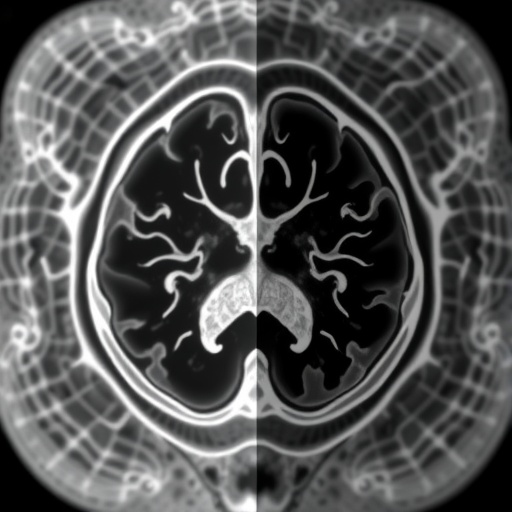
Medical images are rarely as straightforward as they appear on a radiology monitor. Behind every computed tomography scan lies a vast range of pixel intensities, far wider than what the human eye, or a neural network, can meaningfully digest at once. Radiologists have long dealt with this problem using windowing, a technique that remaps the raw pixel values of an image to a narrower display range, amplifying the contrast of the structures that matter. A liver window on a CT scan, for instance, sacrifices detail in bone and lung tissue to make hepatic lesions stand out. Yet while windowing is routine in the clinic, it has remained a curiously neglected corner of artificial intelligence research. A new study published in Biomedical Engineering Letters argues that this oversight may be quietly limiting the accuracy of deep learning systems used to analyze medical scans, and it proposes an elegant fix that lets the machine choose its own windows.
The research, conducted by Jangho Kwon and Kihwan Choi of the Department of Applied Artificial Intelligence at Seoul National University of Science and Technology, introduces a data-driven, multi-contrast windowing method that learns which contrast settings are most useful for a given image analysis task. Rather than relying on hand-picked window widths and levels, the approach trains a neural network module that suggests multiple windows simultaneously, each tuned to the needs of a downstream segmentation model. The result is a pipeline in which the machine not only detects pathology but also reveals, through automatically generated contrast-enhanced images, which parts of the intensity spectrum it considers important for its predictions.
Windowing works by adjusting two parameters: the window width, which controls the dynamic range of displayed intensities, and the window level, which sets the center of that range and therefore the overall brightness. In a CT image, where Hounsfield units span from dense bone to air, a soft-tissue window might compress the display range to roughly 50 to 350 Hounsfield units, making subtle differences within the liver or brain visible. Radiologists have known for decades that this choice is consequential. A 1999 study in Radiology demonstrated that dedicated liver window settings measurably improved the detection of hepatic lesions, and clinical practice has since accumulated an arsenal of preset windows for different organs and pathologies. In magnetic resonance imaging, the challenge is compounded by the multiplicity of pulse sequences, T1-weighted, T2-weighted, and fluid-attenuated inversion recovery images each carry different intensity distributions, and standardization of these scales remains an active research problem in its own right.
When deep learning entered medical imaging, many researchers simply carried over standard clinical windows, or applied simple normalization schemes such as min-max scaling or z-score standardization, without asking whether those choices were optimal for the model. Kwon and Choi argue this is a blind spot. The input transform applied to a network, including windowing, is itself a hyperparameter of the entire system, and a poor choice can obscure precisely the intensity gradients that a segmentation network needs to delineate a tumor’s boundary. Previous efforts have acknowledged the problem: some studies have trained networks on multiple fixed windows and combined their outputs, while others have proposed trainable windowing for specific tasks such as intracranial hemorrhage detection or liver CT segmentation. The new work extends this line of thinking in two significant directions.
The first is the multi-contrast aspect. Instead of committing to a single learned window, the method generates several windowed versions of each input image, each emphasizing a different band of intensities. These multi-contrast images are then fed to subsequent segmentation models, allowing the network to consult complementary views of the same anatomy. The idea echoes how radiologists themselves work, flipping between lung, bone, and soft-tissue windows to build a complete picture. The second contribution is interpretability. Because the learned windows are task-specific, the method can render a contrast-enhanced image that visualizes which windows the downstream model relies on most heavily for its prediction. In other words, the technique produces a kind of window-level attention map, offering clinicians a window into the machine’s decision-making that goes beyond conventional saliency methods such as Grad-CAM.
To achieve this, the authors construct a windowing module that can be inserted into an end-to-end training pipeline and optimized jointly with the segmentation network. The module learns to remap pixel values so that the regions of interest gain contrast at the expense of irrelevant background intensity ranges. The architecture draws on established building blocks from computer vision, including inverted residual structures familiar from MobileNetV2 and squeeze-and-excitation style channel attention, which allow the module to weigh the relative importance of different learned windows dynamically. During training, the whole system is optimized with gradient-based methods so that the windows adapt to whatever the segmentation task demands, whether that is finding a hypodense liver tumor in CT or distinguishing edema from enhancing tumor core in brain MRI.
The experimental evaluation covered three distinct tasks. The first was liver tumor segmentation in CT images, using data drawn from the well-known Liver Tumor Segmentation Benchmark, or LiTS, a widely used community dataset of contrast-enhanced abdominal CT volumes with expert annotations of liver parenchyma and tumors. The second was abdominal organ segmentation in MRI, assessed in the context of the CHAOS combined CT-MR challenge, which tests models on healthy abdominal structures across different modalities. The third was brain tumor segmentation in MRI, a task made notoriously difficult by the heterogeneous intensity signatures of gliomas and the interplay of multiple MRI sequences. Across these benchmarks, the authors compared their multi-contrast windowing against conventional fixed-window preprocessing and against other segmentation backbones, including attention-based U-Net variants, autoencoder-regularized 3D networks, and transformer-based architectures such as Swin UNETR.
The results, according to the study, show consistent gains. Segmentation models that received multi-contrast windowed inputs achieved higher accuracy than the same models fed conventionally windowed images, indicating that the learned windows were indeed capturing intensity information that fixed windows discarded. Just as importantly, the method produced interpretable visual outputs: contrast-enhanced images in which the band of intensities most critical to the model’s decision was emphasized. For a clinician, this means the AI system does not function as an opaque oracle. It effectively communicates, in the visual language of radiology, what it is looking at, an important step for building the trust needed before such systems enter routine diagnostic workflows.
The implications extend beyond the three tasks studied. Segmentation accuracy is a bottleneck for a wide range of clinical applications, from radiation therapy planning, where tumor boundaries determine treatment volumes, to organ-at-risk delineation, surgical navigation, and quantitative imaging biomarkers. If a simple, learnable preprocessing step can meaningfully improve performance without altering the underlying model architecture or requiring additional hardware, it represents an unusually cost-effective upgrade. The method is also modality-agnostic in principle: any imaging pipeline in which the mapping from raw intensity to display value is somewhat arbitrary, whether cone-beam CT, mammography, or microscopy, could in principle benefit from task-specific learned windowing.
There are also subtle scientific insights embedded in the approach. By examining the learned windows across tasks, one can ask whether a network detecting liver tumors converges on windows resembling the clinical liver window, or whether it discovers entirely different intensity bands. The study’s visualization capability makes such questions tractable, potentially informing radiology practice itself: if a machine consistently prefers a certain window for a certain task, that window might reveal contrast relationships that human observers have overlooked, or confirm decades of accumulated radiological wisdom from a new direction.
The work, published online on 13 May 2026 and funded by a research program of Seoul National University of Science and Technology, builds on the authors’ earlier 2020 conference paper on trainable multi-contrast windowing for liver CT segmentation. In the years since, the field has moved toward ever more powerful segmentation architectures, from nnU-Net, a self-configuring framework that dominates many biomedical segmentation leaderboards, to transformer-based models. Yet the preprocessing layer, the humble transformation that determines what the network actually sees, has received comparatively little attention. This study suggests that revisiting that layer with modern deep learning tools can pay dividends that rival changes in architecture.
For the growing community developing AI-based diagnostic tools, the message is clear: the inputs matter as much as the models. A network is only as good as the representation it is fed, and in medical imaging, that representation is shaped long before the first convolutional filter fires. By making windowing itself a learned, task-adaptive, and interpretable component, Kwon and Choi have turned a routine display setting into a source of both accuracy and insight. As deep learning systems move closer to the clinic, techniques like this one, which improve performance while making the machine’s reasoning visually legible to the radiologists who must ultimately trust it, may prove as important as any architectural breakthrough. The study’s approach of learning multi-contrast windows jointly with segmentation models offers a template that other groups can adopt, extend, and test across new modalities, institutions, and disease targets, bringing the field one step closer to medical AI that both sees better and explains itself.
Cite Scienmag News
Blake Davidson. (September 10, 2026). Deep learning enables task-specific multi-contrast medical image visualization. Scienmag. https://scienmag.com/deep-learning-enables-task-specific-multi-contrast-medical-image-visualization/
Blake Davidson. "Deep learning enables task-specific multi-contrast medical image visualization." Scienmag, 10 September 2026, https://scienmag.com/deep-learning-enables-task-specific-multi-contrast-medical-image-visualization/. Accessed 10 September 2026.
Blake Davidson. "Deep learning enables task-specific multi-contrast medical image visualization." Scienmag. September 10, 2026. https://scienmag.com/deep-learning-enables-task-specific-multi-contrast-medical-image-visualization/


