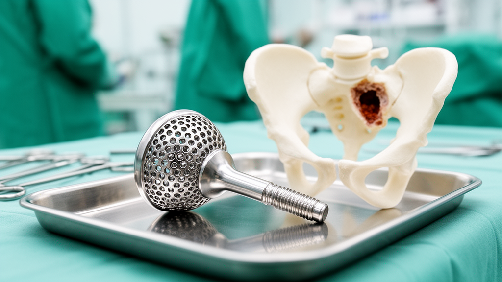
When a hip replacement fails and the bone around it has all but vanished, standard revision implants often have nothing left to grip. A new retrospective study from University Hospital Bonn, published in the journal Arthroplasty, reports 18 years of experience with patient-specific, 3D-printed acetabular implants used in exactly these situations. Across 154 consecutive patients treated between October 2007 and June 2025, the custom-made acetabular components provided mid-term function and implant retention in a cohort whose defects were among the most severe that revision surgeons encounter.
All patients in the study had major acetabular bone loss, classified as Paprosky type 3A in 85 cases and type 3B in 69, corresponding to Acetabular Defect Classification (ADC) type 3C and type 4 defects, the latter defined by pelvic discontinuity, a complete separation of the upper and lower halves of the pelvis. The cohort comprised 48 men (31.2 percent) and 106 women (68.8 percent), with a mean follow-up of 34.82 months, ranging from 6.2 to 168 months. Nearly half of the reconstructions, 74 of 154 patients (48.1 percent), were performed because a previous infection had destroyed the original implant’s foundation, and Staphylococcus epidermidis was the most frequently isolated organism in those cases.
The study’s central narrative is an evolution in implant philosophy. Early reconstructions relied on triflange or biflange constructs, with fixation points anchored into the ilium, pubis, and ischium, a three-point strategy that offered theoretical stability but demanded extensive surgical exposure near the iliac vessels and carried a risk of sciatic nerve injury. Over time, the surgeons observed that osteolysis of the inferior hemipelvis often leaves the pubic and ischial regions too degraded for reliable screw fixation. They progressively abandoned inferior fixation and moved toward a monoflange design, concentrating anchorage in the ilium and the sacroiliac region, where bone stock remains structurally dependable. By the study’s end, 101 patients (65.6 percent) had received monoflange components, 44 (28.6 percent) biflange, and only 9 (5.8 percent) triflange implants.
The current design centers on what the authors call a 90°–90° fixation concept. A 10-mm roughened, macroporous iliac peg is inserted to maximal depth with angular-stable locking, aligned at 90 degrees to the planned acetabular inclination, while cannulated 8-mm trans-iliosacral screws, when used, are positioned at 90 degrees to the iliac flange. In cases with pelvic discontinuity, this cranial-dominant construct deliberately does not attempt to restore bony continuity across the separated pelvis. Instead, it aims to neutralize the discontinuity biomechanically, resisting medial and lateral tilting and counteracting axial, shear, and rotational loads through superior fixation alone. The authors stress that this concept reflects their longitudinal clinical rationale rather than a biomechanically validated principle and remains a hypothesis awaiting direct comparative testing.
Every implant was designed from thin-slice computed tomography obtained after explantation of the failed component, in collaboration with three manufacturers, and produced from medical-grade titanium alloy using additive manufacturing techniques such as electron-beam melting or selective laser melting. The workflow from imaging to implantation takes roughly six to twelve weeks, a delay the authors acknowledge as a practical limitation, since further bone resorption can occur while the patient waits. In recent years, sterile 3D-printed pelvic models have been brought into the operating room to help surgeons confirm implant positioning and screw placement by touch as well as by sight.
Function improved measurably. Among 147 patients with paired scores, the Oxford Hip Score rose from a mean of 21.69 before surgery to 30.93 at the latest follow-up, an improvement of 9.23 points that reached statistical significance with a p value below 0.0001. Because this gain exceeds the minimal important change of 8.6 points reported for revision hip replacement, the authors regard it as clinically meaningful, not merely a statistical artifact. All procedures were staged, with 103 two-stage and 51 multiple-stage reconstructions, reflecting the complexity of the underlying defects.
Survivorship analysis told a more sobering story. With explantation as the endpoint, Kaplan–Meier estimates were 0.85 at three years and 0.78 at five years, falling to 0.62 at the latest follow-up of 168 months. For reoperation from any cause, estimated survival was 0.64 at three years and 0.58 at five years, dropping to 0.42 at 168 months. These late figures rest on very few patients at risk, only 27 at 60 months and 8 at 120 months for explantation, so the long-term estimates carry substantial uncertainty. Nineteen patients ultimately required explantation, nine for periprosthetic joint infection and ten for aseptic or mechanical reasons, and four of those later received a new custom reconstruction.
Complications remained a defining challenge. Postoperative periprosthetic joint infection occurred in 17 patients (11.0 percent) and dislocation in 16 (10.4 percent), making these the two most frequent adverse events. Eight of the infected patients were treated with debridement, antibiotics, and implant retention, while nine required implant removal and a Girdlestone situation. Six of the dislocations were managed successfully with closed reduction alone. Other events included postoperative hematoma in 19 patients (12.3 percent), wound-healing disturbance in 16 (10.4 percent), intraoperative pelvic fractures in 3 cases (1.9 percent), and one sciatic nerve palsy. Notably, dislocation rates were nearly identical whether or not a dual mobility construct was used, 10.4 percent versus 10.3 percent, a difference that was not statistically significant.
An exploratory multivariable Cox model examining implant removal included infection status, pelvic discontinuity, flange design, and age at surgery. Flange design was the only covariate significantly associated with explantation, with a hazard ratio of 17.70 comparing multiflange with monoflange constructs, while pelvic discontinuity, infection status, and age showed no significant association. The authors caution, however, that the small number of events makes this model prone to overfitting, and they treat it strictly as hypothesis-generating rather than proof that monoflange designs outperform multiflange ones. Whether the shift from multiflange to monoflange fixation genuinely translates into better outcomes, they write, will require dedicated comparative studies.
Radiographic results offered grounds for cautious optimism. Among 146 implants available for assessment, 132 (90.4 percent) met the osseointegration criteria of Moore and colleagues, showing at least three of five signs of bony integration at a minimum of twelve months after surgery. Screw breakage was seen in five patients (3.4 percent) and screw malposition requiring revision in two (1.4 percent). The authors emphasize the study’s inherent limits: it was retrospective, single-center, and without a control group, since custom implants are used only when standard reconstruction is impossible. Surgical technique, manufacturing processes, and implant manufacturers all changed over the 18 years, factors inseparably confounded with implant design. Still, for patients facing otherwise untreatable bone loss, the findings suggest that individualized, 3D-printed reconstruction can hold its ground at mid-term follow-up, even as infection and instability continue to test its limits.
Subject of Research: Mid-term clinical and radiographic outcomes of custom-made 3D-printed acetabular implants in severe acetabular bone defects
Article Title: Custom-made acetabular implants for severe acetabular bone defects: implant evolution, mid-term clinical and radiographic outcomes, and complications in 154 consecutive patients over 18 years
Article References: Babasiz, M., Zellner, A. A., Babasiz, T., Ossendorff, R., Fröschen, F. S., Strauss, A. C., Jaenisch, M., & Wirtz, D. C. (2026). Custom-made acetabular implants for severe acetabular bone defects: implant evolution, mid-term clinical and radiographic outcomes, and complications in 154 consecutive patients over 18 years. Arthroplasty, 8(1), Article 65. https://doi.org/10.1186/s42836-026-00425-3
Image Credits: AI Generated
DOI: 10.1186/s42836-026-00425-3
Keywords: custom-made acetabular implants, revision hip arthroplasty, acetabular bone defects, 3D-printed titanium implants, pelvic discontinuity, monoflange fixation, Kaplan-Meier survivorship, Oxford Hip Score, periprosthetic joint infection, dislocation, osseointegration, Paprosky classification
Cite Scienmag News
Ophelia Keating. (October 30, 2026). Custom Hip Implants Show Mid-Term Reliability in Severe Bone Defects. Scienmag. https://scienmag.com/custom-hip-implants-show-mid-term-reliability-in-severe-bone-defects/
Ophelia Keating. "Custom Hip Implants Show Mid-Term Reliability in Severe Bone Defects." Scienmag, 30 October 2026, https://scienmag.com/custom-hip-implants-show-mid-term-reliability-in-severe-bone-defects/. Accessed 30 October 2026.
Ophelia Keating. "Custom Hip Implants Show Mid-Term Reliability in Severe Bone Defects." Scienmag. October 30, 2026. https://scienmag.com/custom-hip-implants-show-mid-term-reliability-in-severe-bone-defects/