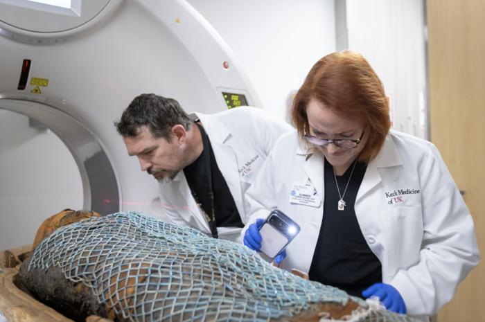
In a groundbreaking fusion of ancient history and cutting-edge medical technology, radiologists at Keck Medicine of the University of Southern California (USC) have employed state-of-the-art computed tomography (CT) scanning to unlock unprecedented insights into the lives of two ancient Egyptian priests. These two mummies, Nes-Min, dating back to approximately 330 BCE, and Nes-Hor, from around 190 BCE, have been preserved within their sarcophagi for over 2,200 years. Using advanced imaging techniques typically reserved for diagnosing modern medical conditions, scientists have revealed intimate details about these individuals’ health, anatomy, and personal histories.
The project harnessed the capabilities of a 320-slice CT scanner, one of the most sophisticated tools available in medical imaging, to conduct full-body scans of the mummies inside the lower halves of their heavy sarcophagi. Such scanners produce hundreds of thin cross-sectional images or “slices” of the body, which digital visualization experts then stack to form a comprehensive three-dimensional digital reconstruction. This digital approach permits an in-depth exploration of the mummies without risking damage to the delicate linen wrappings or the historic remains themselves.
Unlike prior efforts that relied on older scanning technologies, the modern CT scanner’s enhanced resolution has allowed for remarkable detail. The scans revealed facial features unknown until now, such as the contours of their eyelids and the exact shape of their lower lips, providing a more humanized perspective of these long-deceased priests. This breakthrough enables researchers and museum visitors alike to connect with the individuals on a deeply personal level, bridging millennia with the power of medical imaging.
Health assessments gleaned from the scans indicated that Nes-Min likely suffered from chronic lower back pain, a condition common in contemporary populations. His lumbar vertebrae showed signs of collapse consistent with natural wear and tear due to aging, a profound reminder that human physiology has confronted similar ailments across epochs. Additionally, the presence of artifacts buried with Nes-Min, including scarab beetles and fish representations, continued to shed light on burial customs and daily beliefs.
Nes-Hor’s CT images presented a different health profile, exposing severe dental pathology and advanced deterioration of the hip joint. Intriguingly, despite being from a later period, Nes-Hor was evidently older at the time of death than Nes-Min, a fact which underscores the significance of individual life experiences and health challenges faced by ancient peoples. These scans unfold personal health narratives that humanize the past and challenge assumptions about ancient lifespans and medical conditions.
Leading the imaging project, Summer Decker, PhD, the director of the USC Center for Innovation in Medical Visualization, emphasized how advancements in scanning technology have dramatically enhanced the level of detail visible today. “Previous scans could not capture the extensive and detailed information we now have, which opens up new possibilities for understanding these ancient individuals in ways previously unimaginable,” she explained. Her team’s expertise allowed them to transform raw imaging data into vivid 3D models, transcending the limitations of 2D viewing and enhancing interpretative accuracy.
Beyond digital models, the team utilized medical-grade 3D printing technologies to produce life-size replicas of key skeletal elements such as the skulls, spines, and hips of the priests, as well as the artifacts discovered with Nes-Min. These tactile reproductions provide invaluable tools not only for scientific research but also for public exhibition and educational purposes, engaging museum visitors with tangible connections to ancient history.
The “Mummies of the World: The Exhibition” at the California Science Center offers a premier venue for this display. Opening February 7, this exhibit features these newest scanned mummies, bringing never-before-seen detailed digital and physical representations to Los Angeles. According to Diane Perlov, PhD, an anthropologist and senior vice president for special projects at the center, such technological applications offer a “powerful window into the world of ancient people and past civilizations that might otherwise be lost,” enabling a deeper understanding of historical lifeways.
Keck Medicine’s innovations extend far beyond archaeology. Their 3D visualization and printing techniques are pivotal in translating clinical medical imaging—such as CT and MRI—into physical models for surgical planning and education. The process starts with hundreds of cross-sectional slices that are digitally reconstructed into three-dimensional models. Surgeons can then analyze and measure complex anatomical structures with greater precision or create accurate models to rehearse surgeries, improving patient outcomes by providing tailored treatment options based on precise anatomy.
These tangible models also have transformative effects on patient communication. According to Dr. Decker, patients holding replicas of their own organs gain new insights into their medical conditions, fostering understanding and cooperation in their treatment plans. Such technologies bridge the gap between abstract medical imaging and patient experience, demonstrating how advances originally developed for clinical care can reverberate into diverse fields such as archaeology.
The scanning project of Nes-Min and Nes-Hor epitomizes the tremendous interdisciplinary synergy between medical imaging technology, anthropology, and museology. It not only challenges preconceived notions about ancient health and lifestyles but also exemplifies how modern medical technology can stimulate fresh discoveries in humanities research. Access to nearly two dozen 3D printers at the USC Center for Innovation in Medical Visualization underscores Keck Medicine’s commitment to integrating innovation with applied science.
As “Mummies of the World: The Exhibition” showcases these ancient individuals in a new light, it epitomizes the power of technology to contextualize history in a visceral and relatable way. Each scan and print makes a silent millennia-old story visible and palpable, inviting reflection on the enduring human condition. Ultimately, this extraordinary endeavor heralds a future where continued technological advancements will deepen our connection to the past and illuminate its enduring relevance to human health and society.
Subject of Research: Ancient Egyptian Mummies, Medical Imaging, Computed Tomography
Article Title: Unlocking Ancient Lives: 3D CT Scanning Reveals Hidden Stories of Egyptian Mummies
News Publication Date: February 7, 2024
Web References:
Image Credits: Ricardo Carrasco III
Keywords: Imaging, Archaeology, Computed Tomography, 3D Visualization, Mummies, Medical Technology, Ancient Egypt
