


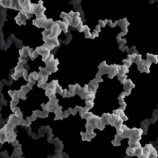
Hereditary transthyretin amyloidosis (hATTR) is driven by the misfolding of transthyretin (TTR), a transport protein whose fragments assemble into toxic amyloid fibrils. In patients, these fibrils accumulate in tissues, underpinning progressive organ dysfunction. Yet, the precise molecular architecture of fibrils formed in living disease—rather than in vitro models—has remained difficult to capture. A new study now addresses that gap by turning to cryo–electron microscopy (cryo-EM) applied directly to biopsy-derived samples.
Using cryo-EM, researchers reconstructed high-resolution fibril structures from patient tissue. They report that TTR fibrils adopt distinct structural arrangements that can be interpreted as disease-relevant “blueprints” for how the protein aggregates. The work provides a framework for understanding how specific hereditary variants influence assembly pathways.
A key technical advance is the ability to resolve fibril polymorphism—variations in how repeated molecular units stack and twist along the fibril axis. Cryo-EM maps reveal ordered segments within the otherwise heterogeneous assembly, enabling the researchers to differentiate conserved core regions from more variable surface features that may affect how fibrils seed new growth.
The study also highlights the role of fibril symmetry and protofilament pairing. Amyloid fibrils are typically built from two intertwined protofilaments, and the authors describe how the relative orientation of these strands shapes β-sheet organization. Such structural details are essential because they dictate fibril stability and the exposure of aggregation-prone surfaces.
By comparing observed patient-derived structures, the team proposes that fibrils can emerge through multiple conformational states rather than a single canonical form. This is consistent with the clinical reality that hATTR shows variable severity across individuals. Understanding which conformations dominate may help explain differences in tissue tropism and progression rate.
Beyond structural description, the findings inform therapeutic strategies aimed at halting fibril formation or promoting clearance. Many interventions rely on interrupting early aggregation steps or blocking elongation surfaces; structure-guided targets can sharpen those approaches. If certain conformers present unique binding pockets or steric constraints, therapies could be tailored accordingly.
Overall, the cryo-EM reconstruction of biopsy-derived TTR fibrils marks a significant step toward translating amyloid biology into actionable molecular models. As more disease-specific fibrils are mapped, the field can better connect genotype, fibril form, and outcomes—turning patient samples into atomic-level evidence.
Cite Scienmag News
Ophelia Keating. (July 28, 2026). Cryo-EM Reveals Biopsy-Derived TTR Fibril Structures in Hereditary Amyloidosis. Scienmag. https://scienmag.com/cryo-em-reveals-biopsy-derived-ttr-fibril-structures-in-hereditary-amyloidosis/
Ophelia Keating. "Cryo-EM Reveals Biopsy-Derived TTR Fibril Structures in Hereditary Amyloidosis." Scienmag, 28 July 2026, https://scienmag.com/cryo-em-reveals-biopsy-derived-ttr-fibril-structures-in-hereditary-amyloidosis/. Accessed 4 September 2026.
Ophelia Keating. "Cryo-EM Reveals Biopsy-Derived TTR Fibril Structures in Hereditary Amyloidosis." Scienmag. July 28, 2026. https://scienmag.com/cryo-em-reveals-biopsy-derived-ttr-fibril-structures-in-hereditary-amyloidosis/


