


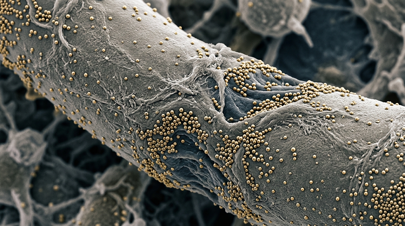
By simulating the physical and electrical properties of these nanoparticles, the team provides quantitative evidence regarding how specific variables affect the efficacy of neural activation.
The central finding of the research is that the stimulation capability of magnetoelectric nanoparticles increases as their concentration within the tissue volume rises. The study further reveals that the random spatial distribution of these particles plays a crucial role in determining the electrical field distribution within the nerve. This variability means that the specific positioning of nanoparticles relative to neural fibers can drastically alter the likelihood of eliciting an action potential, highlighting the importance of stochastic factors in nanoscale neural interfaces.
Magnetoelectric nanoparticles, or MENPs, are composite materials designed to convert magnetic energy into electrical signals. They typically consist of a magnetostrictive core coated with a piezoelectric shell. When exposed to an external magnetic field, the core deforms, and this mechanical strain is transmitted to the shell, which generates an electrical charge. This direct magnetoelectric effect allows for wireless stimulation of neural tissue without the need for invasive electrodes, offering a potential solution to the trade-off between selectivity and invasiveness found in current neural interfaces.
The study employed a holistic computational framework to model this interaction. The researchers used a multiphysics approach to characterize the direct magnetoelectric effect of a single nanorod-shaped nanoparticle. They then scaled this model to simulate multiple nanoparticles distributed randomly within a small volume of nerve tissue. This approach allowed them to assess the electric field distribution generated by the nanoparticles and its subsequent effect on the electrical responses of different types of peripheral axons.
To ensure the models were realistic, the researchers considered the anatomical variability of peripheral nerves. They evaluated the stimulation capability of the nanoparticles against both myelinated and unmyelinated axons, which have profoundly different excitability characteristics. The computational models included eight different peripheral axon types, ranging from small-diameter myelinated fibers to larger myelinated fibers and unmyelinated C-type fibers. This comprehensive approach allowed for a detailed comparison of how different neural dynamics respond to the same stimulation parameters.
The simulations tested three different weight/volume concentrations of nanoparticles: 0.1%, 1%, and 10%. For each concentration, the researchers simulated 50 different random spatial configurations to capture the variability inherent in nanoparticle distribution. The results showed that the median and percentiles of the electric field distribution within the tissue increased with higher nanoparticle concentrations. For instance, the 95th percentile of the electric field values increased from 1.6 × 10^3 V/m at 0.1% concentration to 1.3 × 10^5 V/m at 10% concentration, indicating a substantial enhancement in the electrical environment surrounding the nerve fibers.
A key metric used in the study was the multiplication factor, or MF, which quantifies how much the electric potential generated by the nanoparticles must be amplified to elicit an action potential in a neural fiber. The study found that the required MF values decreased as the nanoparticle concentration increased. Smaller myelinated fibers required higher MF values than larger myelinated fibers, while the relationship between myelinated and unmyelinated fibers varied with concentration. At low concentrations, unmyelinated fibers required lower MFs, but at higher concentrations, myelinated fibers showed greater excitability, likely due to the increased probability of nanoparticles being positioned near the active sites of these fibers.
The probability of eliciting a neural response was also analyzed. At the lowest concentration of 0.1%, the probability of obtaining a multiplication factor below 1, which indicates sufficient stimulation for an action potential, was very low, up to 0.05% for most fiber types. However, at the highest concentration of 10%, the probability increased for larger myelinated fibers, reaching up to 2%. This suggests that while low concentrations may be insufficient for reliable stimulation, higher concentrations can significantly improve the likelihood of neural activation, particularly for more excitable fiber types.
The study also highlighted the importance of the spatial arrangement of nanoparticles. The variability in the electric field distribution was higher at lower concentrations, where the random positioning of fewer particles had a more pronounced effect on the local electric field. As the concentration increased, the variability decreased, suggesting that a higher density of nanoparticles leads to a more consistent and predictable electrical environment. This finding underscores the need for precise control over nanoparticle distribution in future applications to ensure reliable and selective neural stimulation.
Despite the promising results, the study acknowledges several limitations. The nanoparticles were assumed to be distributed within a small, parallelepiped-shaped volume, which limited the analysis to a short length of peripheral fibers. For example, only one Ranvier node was exposed to nanoparticle stimulation in myelinated fibers. This assumption, while useful for characterizing the stimulation capability, may not fully capture the complexity of in vivo conditions. Future studies are needed to develop more realistic models that account for larger tissue volumes and more complex neural geometries.
The implications of this research extend to the development of next-generation neural interfaces. By providing a detailed understanding of how nanoparticle concentration and distribution affect neural stimulation, the study offers valuable insights for the design of minimally invasive, wireless neural devices.
The study by Galletta and colleagues provides a computational framework for evaluating the performance of magnetoelectric nanoparticles in peripheral nerve stimulation.
To capture the underlying physical mechanisms, the researchers modeled each nanorod-shaped device with a cobalt ferrite magnetostrictive core and a barium titanate piezoelectric shell. The nanostructure measured 200 nanometers in height with a 25-nanometer core radius and a 5-nanometer shell thickness, surrounded by tissue with an electrical conductivity of 0.35 siemens per meter and relative permeability of one. When exposed to an external uniform direct-current magnetic field of 100 kiloamperes per meter aligned along its main axis, the core magnetization exceeded 120 kiloamperes per meter. This induced mechanical deformation in the shell, producing a dipole-like surface electric potential between positive and negative 11.5 millivolts, yielding a 22.5-millivolt surface potential difference and a direct magnetoelectric coefficient of approximately 1 volt per centimeter per oersted.
The investigative team placed these nanorods into a simulated tissue volume measuring 1 micrometer by 1 micrometer by 20 micrometers inside a nerve fascicle measuring 12 micrometers by 12 micrometers by 80 micrometers. The tested weight-to-volume concentrations of 0.1 percent, 1 percent, and 10 percent corresponded to exactly 5, 50, and 480 nanoparticles, respectively. Across the 50 simulated configurations generated for each concentration, the median distance between individual particles decreased from 7.3 micrometers at 0.1 percent to 6.1 micrometers at 1 percent and 5.1 micrometers at 10 percent, while maintaining a minimum separation of 0.04 micrometers. Even at the highest concentration of 480 nanoparticles within the small volume, the total particle count remained orders of magnitude below established dose-toxicity limits, which are typically on the scale of billions.
Quantitative evaluation of the field behavior revealed how particle packing governs the local electrical environment. On the outer boundary of the volume facing the adjacent nerve fiber, the average first percentile of the electric field rose from 1.0 volt per meter at 0.1 percent concentration to 1,200 volts per meter at 10 percent, while its coefficient of variability dropped from 91.6 percent to 12.0 percent. The average maximum electric potential registered along the 20-micrometer exposed fiber length increased from 1.2 millivolts at 0.1 percent to 3.8 millivolts at 1 percent and 8.9 millivolts at 10 percent. Concurrently, the proportion of fiber length subjected to potentials exceeding 50 percent of the perceived maximum expanded from 5.0 percent at 0.1 percent concentration to 26.3 percent at 10 percent concentration.
The biological simulations, executed using a 100-hertz sinusoidal stimulus applied over 50 milliseconds, demonstrated distinct recruitment patterns across specific axon models. For small-diameter myelinated fibers modeled via the motor nerve dynamic framework at 1.0 micrometer, the median multiplication factor reached 99 at 0.1 percent concentration and decreased to 5.3 at 10 percent concentration, whereas a 5.7-micrometer myelinated fiber required medians of 370 and 2.0 at those respective concentrations. This requirement for higher gain factors in smaller myelinated axons directly contrasts with Henneman’s traditional size principle. When evaluating an ideal threshold defined as twice the voltage needed to activate a fiber with a single adjacent nanoparticle, unmyelinated fibers targeting the autonomic and somatic systems achieved a 100 percent activation probability at the 10 percent concentration.
Subject of Research: In Silico analysis of concentration and spatial distribution effects of magnetoelectric nanoparticles on peripheral nerve activation
Article Title: In Silico analysis of concentration and spatial distribution effects of magnetoelectric nanoparticles on peripheral nerve activation
Article References: Galletta, V., Bonato, M., Parazzini, M., Chiaramello, E., & Fiocchi, S. (2025). In Silico analysis of concentration and spatial distribution effects of magnetoelectric nanoparticles on peripheral nerve activation. Bioelectronic Medicine, 11(1), Article 25. https://doi.org/10.1186/s42234-025-00187-4
Image Credits: AI Generated
DOI: 10.1186/s42234-025-00187-4
Keywords: Silico, analysis, concentration, spatial, distribution, effects, magnetoelectric, nanoparticles, peripheral, nerve, activation, scientific research
Cite Scienmag News
Denise Maddox. (November 9, 2026). Computational Study Links Nanoparticle Density to Nerve Stimulation. Scienmag. https://scienmag.com/computational-study-links-nanoparticle-density-to-nerve-stimulation/
Denise Maddox. "Computational Study Links Nanoparticle Density to Nerve Stimulation." Scienmag, 9 November 2026, https://scienmag.com/computational-study-links-nanoparticle-density-to-nerve-stimulation/. Accessed 9 November 2026.
Denise Maddox. "Computational Study Links Nanoparticle Density to Nerve Stimulation." Scienmag. November 9, 2026. https://scienmag.com/computational-study-links-nanoparticle-density-to-nerve-stimulation/




