A large multicenter prospective study has reported that combining phacoemulsification with goniosynechialysis, a procedure known as PEI-GSL, achieved durable control of intraocular pressure in patients with primary angle-closure glaucoma who also had cataracts. Among 237 patients who completed twelve months of follow-up, mean intraocular pressure fell from 22.9 plus or minus 10.2 mmHg at baseline to 14.0 plus or minus 3.1 mmHg, an absolute reduction of 8.8 plus or minus 10.1 mmHg, corresponding to a 29.3 percent decrease. The number of antiglaucoma medications dropped from 2.08 plus or minus 1.23 to 0.14 plus or minus 0.52, and the proportion of patients using no medications at all rose from 14.2 percent to 90.3 percent.
The study, published in the journal Eye and Vision, enrolled 289 consecutive patients with primary angle-closure glaucoma and coexisting cataracts who underwent PEI-GSL between November 1, 2022 and March 12, 2024. Participants were recruited from five Chinese eye hospitals: the Eye Hospital of Wenzhou Medical University, the Eye Hospital of Nanchang University, Ningbo Eye Hospital, Dalian Third People’s Hospital, and Tianjin Medical University Eye Hospital. All patients received comprehensive ocular evaluations at baseline and at one week, one month, three months, six months, and twelve months after surgery. Of the 289 enrolled, 237, or 82.0 percent, completed the twelve-month follow-up, with a mean follow-up duration of 12.4 plus or minus 1.8 months.
Primary angle-closure glaucoma is a particularly threatening form of the disease. Glaucoma affected approximately 76 million people worldwide in 2020, with projections indicating 111.8 million by 2040, and primary angle-closure glaucoma accounts for roughly 23.4 million of those cases, nearly half of whom live in China. The risk of blindness associated with this subtype is about three times higher than that of primary open-angle glaucoma, and globally it has blinded approximately 5.25 million people. In this condition, at least 180 degrees of the posterior trabecular meshwork is invisible on gonioscopy, accompanied by glaucomatous optic nerve damage and a matching visual field defect.
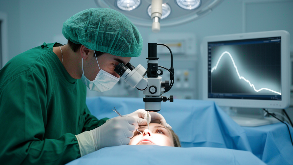
The surgical logic of PEI-GSL rests on two mechanisms. Phacoemulsification removes the cataractous lens and implants an intraocular lens into the capsular bag, which deepens the anterior chamber and can relieve pupillary block. Goniosynechialysis then addresses peripheral anterior synechiae, adhesions between the iris and the trabecular meshwork that block aqueous outflow. In the reported procedure, a senior ophthalmologist made clear corneal and auxiliary incisions of 2.2 to 3.0 millimeters and 1.0 millimeter, filled the anterior chamber with a viscoelastic agent, performed continuous curvilinear capsulorhexis, removed the lens nucleus and cortex by phacoemulsification, and implanted the intraocular lens. The extent of synechiae was assessed intraoperatively by gonioscopy, and viscoelastic and iris repositors were used to separate the adhesions until the trabecular meshwork was fully visualized.
The primary success definition was strict. Complete success required an intraocular pressure of 6 to 18 mmHg without antiglaucoma medications and without reoperation, while qualified success allowed medications but retained the same pressure range and the no-reoperation requirement. By Kaplan-Meier analysis, the complete success rate during follow-up was 80.6 percent and the qualified success rate was 88.3 percent. When the pressure target was relaxed to 6 to 21 mmHg, the corresponding rates rose to 85.5 percent and 94.8 percent. Success rates did not significantly differ between patients with preoperative pressures above or below 21 mmHg, nor between patients using more or fewer than two preoperative medications.
Safety results were also favorable. A total of 56 postoperative complications occurred in 49 patients, or 17.0 percent of the cohort. The most prevalent was corneal edema in 27 patients, or 9.3 percent, followed by postoperative pressure spikes of at least 30 mmHg in 16 patients, or 5.5 percent, hyphema in 6 patients, or 2.1 percent, malignant glaucoma in 3 patients, shallow anterior chamber in 3 patients, and choroidal detachment in 1 patient. All patients with pressure spikes returned to the normal range after antiglaucoma medications. Three patients, or 1.0 percent, underwent yttrium-aluminum-garnet laser posterior capsulotomy because of malignant glaucoma or a shallow anterior chamber, and six patients, or 2.1 percent, required reoperation for uncontrolled pressure or malignant glaucoma, including procedures such as Kahook Dual Blade goniotomy, cyclophotocoagulation, repeat goniosynechialysis, and penetrating canaloplasty.
Notably, the procedure performed comparably in advanced disease. Patients were staged by the mean deviation of their visual fields, with early-to-moderate disease defined as a mean deviation of at least minus 12 decibels and advanced disease as worse than minus 12 decibels. The cohort included 80 patients with early-to-moderate and 209 with advanced glaucoma. Mean pressure fell from 19.7 plus or minus 8.4 mmHg to 14.0 plus or minus 2.6 mmHg in the earlier-stage group and from 23.9 plus or minus 10.6 mmHg to 14.0 plus or minus 3.3 mmHg in the advanced group. Complete success rates were 86.7 percent and 78.2 percent, and qualified success rates were 89.3 percent and 87.9 percent, differences that were not statistically significant. Complication rates, capsulotomy rates, and reoperation rates also did not differ significantly between the groups.
Multivariate Cox regression analysis identified anatomical factors associated with success. Central anterior chamber depth was positively associated with qualified success, with a hazard ratio of 0.25 and a P value of 0.044, while axial length was marginally associated, with a hazard ratio of 0.67 and a P value of 0.050. The authors interpret these associations as reflecting anatomical advantages: a deeper anterior chamber reduces the risk of postoperative angle re-closure and provides a larger potential space between the posterior iris surface and the trabecular meshwork, while a longer axial length is often associated with a more posteriorly positioned lens, reducing the risk of postoperative pupillary block.
The findings strengthen a previously thin evidence base. Earlier studies of PEI-GSL were limited by small samples, early disease stages, or retrospective designs, and patients with advanced glaucoma were excluded from at least one randomized comparison. A prior randomized controlled trial of 38 patients reported complete and qualified success rates of 57.9 percent and 92.1 percent at twelve months with a 27.8 percent pressure reduction, and a retrospective study of 109 patients reported qualified success rates of 85.9 percent and 66.2 percent for pressure targets below 21 and below 18 mmHg, respectively. The new multicenter prospective results, with a larger sample and inclusion of advanced disease, are consistent with and extend those reports, although the authors note their outcomes may partly reflect selection effects, since patients with well-controlled pressures might have been lost to follow-up.
The investigators also acknowledge important limitations. This was a noncomparative observational study without a control arm, so it cannot establish that PEI-GSL outperforms alternatives such as phacotrabeculectomy or phacoemulsification combined with goniosynechialysis and goniotomy, and randomized controlled trials comparing surgical approaches remain warranted. The one-year follow-up is insufficient to judge long-term durability, though the cohort continues to be followed. The sample size may also have been insufficient to detect clinical differences between severity groups, and variations in surgeon expertise and equipment across the five centers might have influenced outcomes. Within those constraints, the study indicates that PEI-GSL substantially lowered intraocular pressure and medication burden with a relatively low incidence of complications, even among patients with advanced primary angle-closure glaucoma and coexisting cataracts.
Subject of Research: One-year outcomes of combined phacoemulsification and goniosynechialysis for primary angle-closure glaucoma with cataracts
Article Title: One-year outcomes of combined phacoemulsification and goniosynechialysis in primary angle-closure glaucoma with cataracts: a multicenter prospective study
Article References: Xiong, K., Li, W., Liu, Q., Zhang, S., Li, G., Mao, H., Huang, Q., Zhang, Q., Le, R., Xie, Y., Zuo, J., Yu, B., Wang, Y., Xu, X., Xing, X., Zhao, L., Yang, L., Liang, Y., & Wang, N. (2026). One-year outcomes of combined phacoemulsification and goniosynechialysis in primary angle-closure glaucoma with cataracts: a multicenter prospective study. Eye and Vision, 13(1), Article 36. https://doi.org/10.1186/s40662-026-00507-y
Image Credits: AI Generated
DOI: 10.1186/s40662-026-00507-y
Keywords: primary angle-closure glaucoma, phacoemulsification, goniosynechialysis, intraocular pressure, cataract surgery, glaucoma surgery, prospective study, surgical outcomes, advanced glaucoma, postoperative complications, antiglaucoma medications, ophthalmology
Cite Scienmag News
Ophelia Keating. (October 29, 2026). Combined Cataract and Glaucoma Surgery Shows Strong One-Year Pressure Control. Scienmag. https://scienmag.com/combined-cataract-and-glaucoma-surgery-shows-strong-one-year-pressure-control/
Ophelia Keating. "Combined Cataract and Glaucoma Surgery Shows Strong One-Year Pressure Control." Scienmag, 29 October 2026, https://scienmag.com/combined-cataract-and-glaucoma-surgery-shows-strong-one-year-pressure-control/. Accessed 29 October 2026.
Ophelia Keating. "Combined Cataract and Glaucoma Surgery Shows Strong One-Year Pressure Control." Scienmag. October 29, 2026. https://scienmag.com/combined-cataract-and-glaucoma-surgery-shows-strong-one-year-pressure-control/