


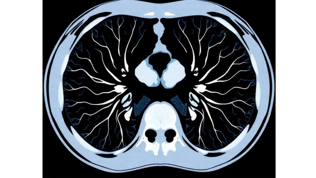
Every chest CT scan carries an invisible enemy: noise. That grainy, mottled texture that overlays the delicate architecture of the lung is not just an aesthetic nuisance—it can obscure the faint differences in density that radiologists rely on to detect early nodules, subtle fibrosis, or the first signs of infection. A new study published in BMC Medical Imaging offers a carefully quantified look at how a modern reconstruction algorithm called KARL 3D, developed for the United Imaging uCT 780 scanner, progressively strips away that noise in lung-window images, and the results paint a striking picture of a dial that radiologists can turn with confidence.
The research, led by Xing Zhang and Yong Peng of the Wuhan Hospital of Traditional Chinese and Western Medicine together with their colleagues, took advantage of an elegant study design known as a within-subject evaluation. Instead of comparing different patients scanned under different conditions—a comparison inevitably muddied by differences in body habitus, lung density, and underlying disease—the team acquired raw CT data from 36 patients once and then reconstructed the very same data multiple times. Each patient’s scan was processed with conventional filtered back projection, the workhorse algorithm of CT for decades, and then again with KARL 3D iterative reconstruction at three escalating strength levels: 3, 6, and 9. Because every reconstruction derived from identical raw data, any difference in image quality could be attributed cleanly to the algorithm itself rather than to biology or chance.
The technical setup matters for understanding what the team measured. All scans were performed at 120 kilovoltage peak with a mean volume CT dose index of 12.0 milligrays, a routine chest protocol. The lung window is notoriously the harshest test for any reconstruction algorithm, because the sharp, edge-enhancing kernels used to visualize lung parenchyma inherently amplify noise. The researchers placed a small circular region of interest, roughly 20 square millimeters, in the lung parenchyma just behind the right main bronchus at the level of the carina, deliberately avoiding vessels and airways. Within that tiny patch of tissue, they measured the standard deviation of pixel values in Hounsfield units—the standard, if imperfect, proxy for image noise—and calculated the signal-to-noise ratio.
The numbers tell a dramatic story. With filtered back projection, lung parenchyma noise averaged 51.76 Hounsfield units, with a standard deviation across patients of 13.31. At KARL 3D level 9, the strongest setting tested, that figure fell to 33.71 Hounsfield units—a reduction of nearly 35 percent. Signal-to-noise ratio climbed in lockstep, from 18.17 to 27.61. Crucially, every pairwise comparison between reconstruction settings reached statistical significance, and the repeated-measures analysis of variance produced an F statistic of 338.7 for the noise measurements, an extraordinarily large value that reflects how consistently the algorithm’s effect manifested across all 36 subjects. The denoising was not a subtle statistical whisper; it was a loud, graded, monotonic effect that strengthened with each step up the reconstruction ladder.
Objective metrics alone, however, do not decide what radiologists actually see. A reconstruction can lower noise on paper while smearing away the fine reticular detail of the lung interstitium, trading one diagnostic liability for another. To address this, two radiologists independently reviewed the image sets and scored them on a five-point Likert scale across four dimensions: perceived noise, structural sharpness, artifact burden, and overall diagnostic acceptability. The composite subjective score rose from 3.21 with filtered back projection to 4.68 at the highest KARL 3D level, indicating that human observers, not just pixel statistics, registered the improvement. Agreement between and within the readers was assessed rigorously: intraclass correlation coefficients for the objective noise measurements ranged from 0.970 to 0.983, and quadratic-weighted kappa values for the subjective summed scores reached 0.86 to 0.88 between readers and 0.79 to 0.96 within the same reader across sessions—figures that fall in the good-to-excellent range and lend credibility to the perceptual findings.
One of the study’s most clinically consequential questions concerned body size. Radiology has long known that larger patients absorb more X-rays, produce noisier images, and often benefit differently from noise-reduction algorithms. If iterative reconstruction’s benefit shrank in larger patients, protocols might need to be tailored by body habitus, complicating workflow. The researchers therefore computed each patient’s effective diameter and tested whether it correlated with the degree of noise reduction. It did not. Pearson correlation coefficients hovered between 0.07 and 0.14, all statistically insignificant, and subgroup comparisons using the Mann-Whitney U test found no difference between body-size groups, with P values between 0.70 and 0.75. Within the range of patient sizes studied, the denoising effect of KARL 3D proved remarkably uniform.
That uniformity has practical implications. It suggests that a single reconstruction level can plausibly serve a broad patient population without per-patient adjustment, simplifying protocol design and reducing one more source of variability in a field already crowded with scanner models, kernel choices, and window settings. It also strengthens the argument for within-subject designs in imaging research more broadly: by holding anatomy and dose constant, the study isolated the algorithm’s contribution with a precision that cross-sectional comparisons rarely achieve.
The study is not without boundaries worth acknowledging. Thirty-six patients constitute a modest sample, all scanned on a single scanner model at a single institution with one dose level and one lung-window kernel. The retrospective design, approved by the institutional ethics committee with a waiver of informed consent, means the findings describe what the algorithm does under one specific set of conditions rather than guaranteeing identical behavior elsewhere. The authors themselves frame the conclusion carefully: KARL 3D produces a graded, level-dependent reduction in lung-window noise, and the choice of level should be guided by the clinical task. That last clause is the quiet heart of the paper. Higher levels progressively cleaned the images and improved subjective quality, but the optimal setting for detecting a subtle ground-glass opacity may differ from the optimal setting for evaluating a known nodule’s margins, and the study provides the gradient map rather than a single prescription.
The broader context makes the work timely. Iterative reconstruction has swept through clinical CT over the past decade as vendors raced to deliver lower radiation doses without sacrificing image quality, replacing filtered back projection in many departments. Yet the proliferation of proprietary algorithms—each with its own strength scale, its own noise texture, and its own trade-offs between smoothness and resolution—has left radiologists navigating a patchwork of incomparable settings. Studies that rigorously characterize one algorithm’s dose-response curve, using objective measurements and blinded observer scores with validated agreement statistics, are the raw material from which evidence-based protocol decisions are built. This paper adds a well-documented data point for a widely deployed Chinese scanner platform, where such characterization has been comparatively sparse.
For patients, the significance is indirect but real. Cleaner lung-window images at a fixed radiation dose mean radiologists can read chest CTs with less of the visual static that breeds uncertainty, and the demonstrated insensitivity to body size means that benefit extends across the patient spectrum rather than concentrating in the slim. For the imaging community, the message is that the KARL 3D dial behaves predictably: turn it up, and noise falls in a graded, statistically robust fashion, with observer agreement confirming that what the numbers promise is what the eyes receive. The next chapter—mapping which levels best serve which clinical questions, across more scanners, doses, and patient cohorts—remains open, but this study has drawn the baseline gradient with unusual clarity.
Subject of Research: Effect of KARL 3D iterative reconstruction levels on image noise in lung-window chest CT
Article Title: Gradient effect of KARL 3D iterative reconstruction on image noise in lung-window chest CT: a within-subject evaluation
Article References: Gradient effect of KARL 3D iterative reconstruction on image noise in lung-window chest CT: a within-subject evaluation. (n.d.). https://doi.org/10.1186/s12880-026-02922-y
Image Credits: AI Generated
DOI: 10.1186/s12880-026-02922-y
Keywords: KARL 3D, iterative reconstruction, chest CT, lung window, image noise, signal-to-noise ratio, filtered back projection, effective diameter, radiology, medical imaging, computed tomography, image quality
Cite Scienmag News
Ophelia Keating. (October 14, 2026). Chest CT Scans Get Sharper and Cleaner as Iterative Reconstruction Levels Rise. Scienmag. https://scienmag.com/chest-ct-scans-get-sharper-and-cleaner-as-iterative-reconstruction-levels-rise/
Ophelia Keating. "Chest CT Scans Get Sharper and Cleaner as Iterative Reconstruction Levels Rise." Scienmag, 14 October 2026, https://scienmag.com/chest-ct-scans-get-sharper-and-cleaner-as-iterative-reconstruction-levels-rise/. Accessed 14 October 2026.
Ophelia Keating. "Chest CT Scans Get Sharper and Cleaner as Iterative Reconstruction Levels Rise." Scienmag. October 14, 2026. https://scienmag.com/chest-ct-scans-get-sharper-and-cleaner-as-iterative-reconstruction-levels-rise/


