


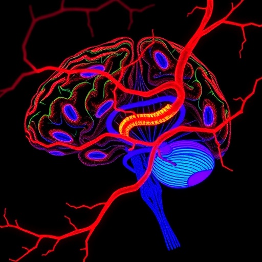
A previously underappreciated protein in the cells lining blood vessels may hold the key to how the brain builds and maintains its protective vascular system. Researchers have found that CD98 heavy chain, commonly known as CD98hc, SLC3A2 or 4F2hc, is essential for the development of blood vessels in the central nervous system (CNS) and for preserving the blood–brain barrier (BBB) throughout life. The study, published in Nature Cardiovascular Research, reveals that the protein has a specialized role in CNS endothelial cells, the cells that form the inner lining of brain and spinal cord blood vessels. When CD98hc was removed from these cells in mice, the animals developed abnormal brain angiogenesis, defective barrier formation and cerebral hemorrhage during embryonic development. In adult mice, the same molecular defect caused breakdown of the BBB and neurological problems, even though the animals did not show a major loss of normal CNS vessel growth.
The findings are significant because the brain’s vascular network is not simply a smaller version of the circulation found elsewhere in the body. CNS blood vessels must deliver oxygen and nutrients while tightly restricting the movement of potentially harmful substances from the bloodstream into neural tissue. This selective interface depends on highly specialized endothelial cells, which form unusually tight cell–cell junctions and coordinate with neurons, glial cells and surrounding vascular support cells. The BBB is therefore both a transport system and a defense system. Its failure has been linked to stroke, neurodegeneration, inflammation, seizures and other neurological disorders. Yet the molecular mechanisms that make CNS endothelial cells different from endothelial cells in peripheral organs have remained only partly understood. The new work identifies CD98hc as one of the proteins that gives the CNS vasculature its distinctive behavior.
Using mouse models and comparisons with human vascular tissue, the investigators found that CD98hc is selectively enriched in CNS endothelial cells compared with endothelial cells from peripheral blood vessels. This pattern suggests that the protein is not merely a general component of the vascular system, but is particularly important in the brain and spinal cord. Such selective enrichment may help explain why endothelial cells in different organs respond differently to the same growth signals or environmental stresses. The discovery also offers a molecular foothold for investigating why the BBB is uniquely vulnerable in some diseases and why treatments that influence blood vessels throughout the body may have very different effects inside the CNS. The presence of CD98hc in human CNS endothelium further raises the possibility that the mechanism could be relevant to human cerebrovascular biology rather than being limited to laboratory mice.
To test its function, the researchers genetically ablated CD98hc specifically in endothelial cells during embryonic development. The consequences were striking but regionally selective. Developing CNS vessels grew in an aberrant pattern, the BBB failed to form properly and blood leaked into brain tissue, producing cerebral hemorrhage. By contrast, the peripheral vasculature was not comparably disrupted. This distinction indicates that CD98hc is not universally required for endothelial survival or vessel formation. Instead, its loss appears to expose a special dependency of CNS endothelial cells, which must coordinate angiogenesis—the growth and remodeling of blood vessels—with the establishment of a tightly sealed barrier. In the developing brain, those processes are closely intertwined: vessels must reach the correct regions, acquire specialized properties and prevent uncontrolled leakage as neural tissue matures.
The researchers also examined what happens after development, when the CNS vascular network has already been established. In adult mice lacking endothelial CD98hc, routine CNS angiogenesis remained largely intact under homeostatic conditions. That result separates two functions that are often considered together: building the vascular network and maintaining its barrier properties. The mature vessels did not simply disappear, and the animals did not show evidence that normal vessel maintenance depended on continuous CD98hc-driven angiogenesis. Instead, the primary defect was a weakened BBB, accompanied by neurological deficits. The observation suggests that CD98hc has a continuing role in the functional stability of adult brain vessels, even after its developmental role in shaping the vascular network has diminished.
The mechanism identified by the study centers on the integrin–FAK signaling pathway. Integrins are cell-surface receptors that allow endothelial cells to sense and attach to the surrounding extracellular matrix, the structural material that supports tissues. When integrins are engaged, they can activate focal adhesion kinase, or FAK, an intracellular signaling protein that coordinates adhesion, cytoskeletal organization, cell movement and communication with growth-factor receptors. According to the researchers, loss of endothelial CD98hc produces a CNS-specific reduction in this systemic integrin–FAK pathway. The weakened signal then affects downstream vascular programs involving VEGFR2 and Wnt–β-catenin, two pathways that are central to vessel growth, endothelial specialization and BBB formation.
VEGFR2 is a major receptor for vascular endothelial growth factor, a powerful regulator of angiogenesis. Its activity must be precisely controlled: insufficient signaling can impair vessel growth, while excessive or poorly coordinated signaling can produce abnormal, leaky vessels. The Wnt–β-catenin pathway is equally important in the CNS vasculature, where it helps instruct endothelial cells to adopt BBB characteristics. β-catenin can influence gene expression after receiving signals through Wnt receptors, helping regulate the production of proteins involved in endothelial identity and barrier integrity. The study places the integrin–FAK system upstream of these pathways in the CD98hc-deficient setting, suggesting that CD98hc helps endothelial cells translate their physical interaction with the surrounding matrix into the molecular instructions required for proper CNS vascular function.
One of the most important experiments tested whether the defect could be reversed rather than merely described. When the researchers activated FAK in CD98hc-deficient mice, the CNS vascular phenotype was fully rectified, according to the study. This rescue experiment provides stronger evidence for a causal relationship between CD98hc and the integrin–FAK pathway. Rather than acting as an unrelated marker of endothelial dysfunction, CD98hc appears to support a signaling circuit whose failure triggers the downstream abnormalities in VEGFR2 and Wnt–β-catenin activity. The result also identifies FAK as a potential therapeutic entry point. If the pathway can be manipulated safely and selectively, it may be possible to strengthen the BBB or correct vascular defects without broadly stimulating or suppressing blood vessels throughout the body.
The findings could influence how scientists approach cerebrovascular disease, but they also highlight the challenge of targeting the brain’s circulation without disturbing the rest of the vascular system. Because CD98hc is enriched in CNS endothelium and its loss has relatively limited effects on peripheral vasculature in the reported models, therapies aimed at this molecular axis might offer a degree of anatomical selectivity. That possibility is still speculative, and the study does not establish a treatment for human disease. FAK is involved in many biological processes, including cell adhesion and tissue repair, so systemic manipulation could carry substantial risks. Future studies will need to determine how CD98hc is regulated, whether its activity changes during stroke or neuroinflammation, and whether the same pathway contributes to BBB disruption in human patients.
For now, the work provides a new explanation for how brain blood vessels acquire and preserve their specialized identity. CD98hc appears to function as a CNS endothelial regulator that connects the cell’s external environment to the signaling networks controlling angiogenesis and barrier integrity. Its importance changes with age: during embryonic development, it is required for correctly patterned CNS angiogenesis and BBB formation; in adulthood, it is primarily needed to maintain the barrier and neurological function. By showing that direct FAK activation can restore the vascular phenotype in deficient mice, Hu and colleagues have moved beyond identifying a correlation and toward defining a potentially actionable mechanism. The study opens a promising route for developing therapies designed not simply to alter blood vessels, but to repair the unique vascular interface that protects the brain.
Cite Scienmag News
Cassandra Pierce. (August 24, 2026). CD98hc Controls Brain Angiogenesis and Blood–Brain Barrier Integrity via Localized Integrin–FAK Signaling. Scienmag. https://scienmag.com/cd98hc-controls-brain-angiogenesis-and-blood-brain-barrier-integrity-via-localized-integrin-fak-signaling/
Cassandra Pierce. "CD98hc Controls Brain Angiogenesis and Blood–Brain Barrier Integrity via Localized Integrin–FAK Signaling." Scienmag, 24 August 2026, https://scienmag.com/cd98hc-controls-brain-angiogenesis-and-blood-brain-barrier-integrity-via-localized-integrin-fak-signaling/. Accessed 4 September 2026.
Cassandra Pierce. "CD98hc Controls Brain Angiogenesis and Blood–Brain Barrier Integrity via Localized Integrin–FAK Signaling." Scienmag. August 24, 2026. https://scienmag.com/cd98hc-controls-brain-angiogenesis-and-blood-brain-barrier-integrity-via-localized-integrin-fak-signaling/


