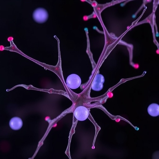
In the complex world of cellular biology, capturing the fleeting moments of rapid intracellular processes has long posed a formidable challenge to scientists. Optical microscopy, a cornerstone technique for investigating live cells, traditionally wrestles with a fundamental trade-off between temporal resolution and image quality. High-speed events often blur or vanish entirely in noisy, photon-starved images, hampering researchers’ ability to visualize biological dynamics with both clarity and precision. However, a pioneering breakthrough from The University of Osaka promises to revolutionize this landscape, unveiling a cutting-edge cryo-optical microscopy method that freezes cellular moments in time with unprecedented spatial and temporal fidelity.
The groundbreaking study, recently published in Light: Science & Applications, details how Osaka researchers have ingeniously combined rapid cryo-fixation with advanced optical imaging to capture snapshotted cellular states previously unattainable with live-cell microscopy alone. By physically arresting biological events mid-motion through instantaneous freezing under the microscope, scientists can now observe dynamic processes not as fleeting blurbs of movement but as pristinely preserved, high-resolution stills. This paradigm shift circumvents long-standing limitations inherent to conventional live-cell imaging, enabling a powerful synthesis of temporal “arrest” and spatial detail.
To achieve this, the research team engineered an avant-garde sample-freezing chamber adjacent to an optical microscope. Unlike traditional cryo-techniques that often involve post-fixation imaging, this approach facilitates rapid vitrification of live cells in situ, effectively “pausing” their internal dynamics precisely when desired. This instantaneous sample immobilization unlocks the capacity for diverse imaging modalities, including super-resolution techniques typically constrained by their slow acquisition rates. By turning the microscope into a temporal freeze-frame camera, the investigators harness the strengths of both live observation and cryo-preservation.
One compelling demonstration involved capturing the rapid propagation of intracellular calcium ion waves within live cardiomyocytes—heart muscle cells—key physiological drivers that orchestrate cellular excitation and contraction. These calcium transients are notoriously difficult to observe in real time due to their speed and subtle fluorescence signals. Using their cryo-optical system, the team successfully froze the calcium wavefront, subsequently applying three-dimensional super-resolution microscopy to reveal intricate structural characteristics of calcium signaling domains at an unprecedented level of detail. This marriage of temporal freezing and enhanced spatial resolution represents a critical advance in decoding the mechanisms of cellular physiology.
Crucially, the methodology does not merely snapshot static images but preserves quantitative information with high fidelity. The extended exposure times enabled by freezing cells with fluorescent calcium indicators allow the collection of far more photons than fleeting live-cell imaging permits. This results in dramatically improved signal-to-noise ratios and quantitative accuracy in measuring intracellular concentrations and dynamics. Researchers are now empowered to conduct precise, reproducible analyses of transient biochemical events that were previously obscured by photonic limitations.
Achieving such temporal precision required ingeniously integrating an electrically triggered cryogen injection system capable of freezing samples within milliseconds of stimulation onset. In experiments inducing calcium waves through UV light, this setup enabled freezing at user-defined timepoints with remarkable 10 ms accuracy. By synchronizing cryo-triggering with biological stimulation, the team could arrest cellular processes at narrowly defined phases, peeling back layers of temporal complexity underlying rapid signaling cascades and transient biochemical states.
The benefits extend beyond a single imaging modality. By instantly halting cellular activity, multiple microscopy techniques can be sequentially applied to the same sample without temporal mismatch artifacts. In a striking showcase, the researchers combined spontaneous Raman microscopy—which yields label-free chemical information—with super-resolution fluorescence imaging on identical frozen specimens. This multimodal approach affords a multidimensional perspective on the same cellular snapshot, marrying molecular composition and structural detail in a way previously impossible for fast biological phenomena.
This innovation heralds a new era in microscopy, particularly for life sciences and biomedical research reliant on accurate visualization of dynamic processes. The ability to “freeze” and then analyze transient events with nanoscale resolution, coupled with versatile imaging modalities, opens vast opportunities to unravel mechanisms behind rapid physiological changes, disease progression, and cellular responses to external stimuli. With scalable potential, this cryo-optical platform promises to become an indispensable tool in the armory of cell biologists and medical researchers alike.
The fundamental concept underpinning this technique—shifting focus from chasing speed to immobilizing dynamics—represents a strategic philosophical leap. Instead of attempting to capture high-speed cellular events in real time and struggling against photon limitations, researchers arrest the biological motion altogether, trading temporal continuity for temporal precision. This shift not only enhances image quality and quantification reliability but also ultimately enriches biological insight by revealing the “still frames” composing complex life processes.
Backing this approach is a synthesis of optical engineering, cryogenic technology, and biological insight, showcasing interdisciplinary innovation at its finest. The team’s results attest to the practicality of integrating cryo-fixation into optical workflows, paving the way for widespread adoption. Moreover, by preserving live-cell spatial and temporal information at the moment of freezing, the approach retains biological relevance typically lost in conventional cryo-based preparations.
In summary, The University of Osaka’s time-deterministic cryo-optical microscopy delivers a transformative tool that enables scientists to freeze rapid intracellular dynamics and analyze them post-fixation with exceptional spatial and temporal precision. This dual advantage overcomes long-standing imaging trade-offs and expands horizons for multimodal, high-fidelity biological investigation. As researchers continue to probe the energetic and fleeting inner workings of cells, this technique equips them with a persuasive new lens through which to witness the choreography of life itself.
Subject of Research: Cells
Article Title: Time-deterministic cryo-optical microscopy
News Publication Date: 23-Aug-2025
References: DOI: 10.1038/s41377-025-01941-8
Image Credits: 2025, Kosuke Tsuji, Masahito Yamanaka et al., Time-deterministic cryo-optical microscopy, Light: Science & Applications
Keywords: Optical microscopy, Fluorescence microscopy, Structured illumination microscopy, Live cell imaging, Cardiomyocytes, HeLa cells, Calcium imaging, Organelles, Live cells, Super resolution imaging


