


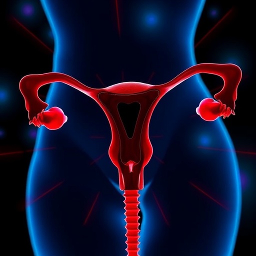
A rare ovarian tumor that can transform the body with male-pattern hormonal changes may be compatible with fertility preservation in carefully selected patients, according to a single-center study from China. The research, conducted at Peking Union Medical College Hospital, examined the cancer and reproductive outcomes of 30 patients with ovarian steroid cell tumors, an uncommon group of ovarian neoplasms whose cells can produce steroid hormones. The findings suggest that patients whose tumors were confined to one ovary and classified as FIGO stage IA generally had favorable cancer outcomes, including no observed recurrences during follow-up. But the study also delivers a sharp warning: fertility-sparing surgery became considerably more uncertain when tumors had ruptured, spread beyond the ovary, or were diagnosed at stage IC or stage II. The work offers one of the most detailed looks yet at the difficult balance between treating a rare ovarian cancer and protecting a patient’s future reproductive options.
Steroid cell tumors are unusual because their biology is defined not only by where they grow, but also by the hormones they can manufacture. They arise from ovarian cells capable of producing steroid hormones, including testosterone and other androgens. When a tumor releases substantial amounts of testosterone, a patient may develop virilizing symptoms: increased facial or body hair, acne, deepening of the voice, male-pattern hair loss, menstrual disruption, or enlargement of the clitoris. These changes can appear gradually and may be mistaken for more common endocrine conditions, such as polycystic ovary syndrome. In the new study, elevated serum testosterone accompanied by virilizing manifestations was present in all 19 patients who underwent fertility-sparing surgery. It was also found in five of the 11 patients who underwent more extensive, non-fertility-sparing procedures. This hormonal signature can provide an important diagnostic clue, although not every steroid cell tumor produces enough hormones to cause obvious symptoms.
The investigators retrospectively reviewed patients who underwent surgery at the Beijing hospital between January 2008 and August 2025. They divided the group according to whether surgeons attempted fertility-sparing surgery, meaning that the uterus and at least part of the reproductive potential were retained. Nineteen patients were placed in the fertility-sparing group, while 11 underwent non-fertility-sparing surgery. The age difference between the groups was striking. Patients receiving fertility-sparing treatment had a median age of 27 years, with ages ranging from 4 to 42. The non-fertility-sparing group had a median age of 55, ranging from 25 to 80. That contrast reflects both medical decision-making and the realities of ovarian cancer care: younger patients are more likely to seek preservation of fertility, while older patients may have completed childbearing or face different surgical considerations. It also means the two groups cannot be interpreted as directly comparable treatment arms in a clinical trial.
Cancer stage emerged as one of the strongest signals in the analysis. The International Federation of Gynecology and Obstetrics, or FIGO, classifies ovarian cancer according to how far it has spread. Stage IA generally indicates that the tumor is limited to a single ovary, with the ovarian capsule intact and no malignant cells detected in relevant fluid or washings. In the fertility-sparing group, 12 of 19 patients, or 63.2 percent, had stage IA disease. Five patients, representing 26.3 percent, had stage IC1 disease, while two patients, or 10.5 percent, had stage II disease. Stage IC1 typically involves surgical spill or rupture of the tumor, whereas stage II indicates extension to pelvic structures. Every patient in the non-fertility-sparing group had stage IA disease, a distribution that highlights the complexity of the surgical choices. Despite the apparent severity of some cases in the fertility-sparing group, no patient with stage IA disease experienced a recurrence during the study’s follow-up period.
The less reassuring results appeared among patients with disease beyond stage IA. Recurrence developed in one patient with stage IC disease who had undergone ovarian cystectomy, an operation that removes the tumor or cyst while leaving as much ovarian tissue as possible. Two patients with stage II disease in the fertility-sparing group also experienced recurrence, and one of them died from the disease. These outcomes do not prove that fertility-sparing surgery caused the recurrences; patients with stage IC or stage II tumors already had biological or anatomical features associated with greater risk. But they do show why a decision to preserve reproductive organs cannot be based on age or fertility preference alone. Tumor stage, capsule status, surgical spill, histological features, and the completeness of tumor removal all matter. In a rare cancer for which evidence is drawn largely from small retrospective series, even a few events can substantially influence clinical interpretation.
The surgical approach considered most reasonable for selected patients with stage IA disease was unilateral salpingo-oophorectomy. This procedure removes one ovary and its associated fallopian tube while leaving the opposite ovary and the uterus intact. In principle, the remaining ovary can continue releasing eggs and producing reproductive hormones, and the uterus remains available for pregnancy. The study’s findings support considering this approach when a tumor is restricted to one ovary and the patient strongly desires future fertility. By contrast, fertility preservation in stage IC or stage II disease should be individualized with caution, the authors concluded. Removing only the tumor through cystectomy may preserve more ovarian tissue, but it can also raise concerns about incomplete excision, tumor rupture, or microscopic disease left behind. The balance between oncological safety and reproductive preservation therefore requires multidisciplinary assessment rather than a one-size-fits-all rule.
The reproductive outcomes in the cohort were far less definitive than the cancer outcomes. At the last follow-up, only one patient clearly reported an active desire for childbearing, and that patient subsequently achieved a spontaneous twin pregnancy that resulted in live birth. This is an encouraging result, but it cannot be interpreted as evidence that fertility-sparing surgery reliably restores fertility after treatment. The small number of patients attempting pregnancy makes it impossible to calculate a meaningful pregnancy rate, and the study did not establish how many participants were infertile, had no pregnancy plans, were too young to attempt conception, or faced other reproductive barriers. The ovarian reserve—the remaining supply of eggs—may be affected by surgery, age, the underlying tumor, or other medical factors. Hormonal recovery was assessed as part of the study, but the available results do not establish a predictable pathway from removal of a steroid cell tumor to successful conception.
The patients were followed for a median of 61 months in the fertility-sparing group and 67 months in the non-fertility-sparing group. The ranges extended as long as 184 months and 160 months, respectively, providing an opportunity to detect some late recurrences. Long-term surveillance is particularly important for steroid cell tumors because their malignant potential is difficult to predict. Histological appearance alone may not always capture the behavior of an individual tumor, and recurrence can depend on factors such as size, necrosis, hemorrhage, atypical cellular features, mitotic activity, and extra-ovarian spread. Follow-up may include clinical examinations, imaging, and measurement of hormones such as testosterone when they were elevated before treatment. A return of virilizing symptoms or a renewed rise in androgen levels could signal recurrent hormone-producing tissue, although hormonal markers must be interpreted alongside imaging and pathology rather than used in isolation.
The study’s design places important limits on what can be concluded. It was retrospective, meaning that the researchers analyzed existing medical records rather than assigning patients prospectively to different treatments. All 30 patients came from a single specialized hospital, which may see unusually complex cases and may have surgical expertise not available everywhere. The fertility-sparing and non-fertility-sparing groups also differed greatly in age and clinical context, making direct comparisons vulnerable to confounding. Most importantly, ovarian steroid cell tumors are so rare that even a carefully assembled cohort remains small. The authors therefore call for multi-institutional studies that combine cases across hospitals and countries, use standardized pathological criteria, track ovarian reserve and menstrual recovery, and record pregnancy attempts systematically. For now, the evidence points to a cautiously hopeful message: stage IA disease may allow preservation of reproductive potential without an obvious compromise in cancer control, but patients with stage IC or stage II tumors face a far more precarious trade-off that must be decided case by case.
Cite Scienmag News
Nathaniel Bowman. (August 27, 2026). Cancer and Fertility Outcomes in Patients With Ovarian Steroid Cell Tumors. Scienmag. https://scienmag.com/cancer-and-fertility-outcomes-in-patients-with-ovarian-steroid-cell-tumors/
Nathaniel Bowman. "Cancer and Fertility Outcomes in Patients With Ovarian Steroid Cell Tumors." Scienmag, 27 August 2026, https://scienmag.com/cancer-and-fertility-outcomes-in-patients-with-ovarian-steroid-cell-tumors/. Accessed 3 September 2026.
Nathaniel Bowman. "Cancer and Fertility Outcomes in Patients With Ovarian Steroid Cell Tumors." Scienmag. August 27, 2026. https://scienmag.com/cancer-and-fertility-outcomes-in-patients-with-ovarian-steroid-cell-tumors/


