


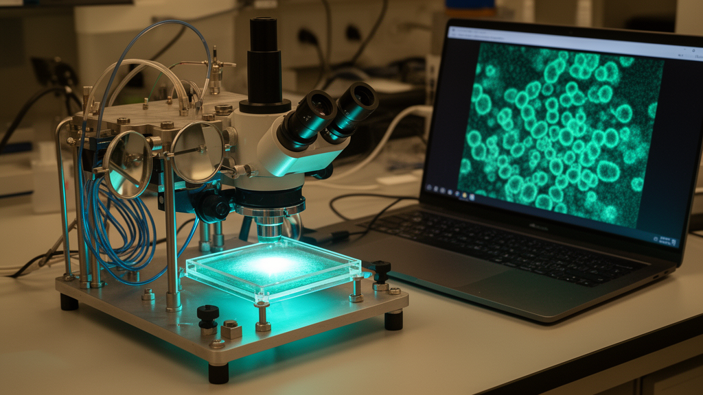
For most life sciences laboratories, the dream of watching individual molecules inside cells with nanometer-scale precision has been locked behind a financial and technical barrier. The most powerful single-molecule localization microscopy techniques, capable of resolving features smaller than ten nanometers, have typically demanded dedicated, expensive hardware: stabilized laser tables, precision optical mounts, active focus-locking systems, and total internal reflection fluorescence (TIRF) illumination modules that few standard facilities can afford. Now, a team of researchers from Uruguay, Argentina, Switzerland, and Germany reports in Communications Engineering a cost-effective and straightforward recipe for converting an ordinary commercial epifluorescence microscope into a high-performance platform that rivals purpose-built instruments for sub-10-nanometer DNA-PAINT and (d)STORM imaging.
The significance of the work lies in what it democratizes. DNA-PAINT, which stands for DNA Point Accumulation for Imaging in Nanoscale Topography, has emerged as one of the most conceptually elegant super-resolution methods available. Instead of relying on photoswitchable fluorescent proteins or dyes, it exploits the transient binding of short, dye-labeled DNA imager strands to complementary docking strands attached to the target molecule. Each binding event produces a brief flash of fluorescence, and because the strands bind and unbind repeatedly, the same target can blink thousands of times. When thousands of these stochastic flashes are localized and summed computationally, the resulting image achieves lateral localization precision better than five nanometers across fields of view as large as 100 by 100 square micrometers, and the approach can multiplex up to thirty different targets simply by changing DNA sequences.
But DNA-PAINT has a demanding appetite for optical stability. Because the technique achieves its extraordinary precision by pinpointing the center of each single-molecule flash, any drift of the sample relative to the objective lens during a minutes-long acquisition translates directly into blurred reconstructions. At the nanometer scale, even the thermal expansion of a microscope body or the slow sag of a stage can ruin an experiment. This is why the gold-standard implementations of DNA-PAINT have relied on TIRF illumination, which confines excitation light to a thin evanescent field near the coverslip surface, dramatically reducing out-of-focus background and boosting the signal-to-noise ratio of individual molecule flashes. Building such a system from scratch, with active focus stabilization, has remained complex and costly for the majority of laboratories.
The new study tackles this bottleneck head-on. The researchers started with a standard commercial epifluorescence microscope of the kind found in countless light microscopy facilities around the world, and they designed a conversion pathway that upgrades it into a stable TIRF platform suitable for single-molecule work. Rather than requiring users to rebuild the microscope’s optical path from raw components, the approach provides a practical engineering route that a competent facility can implement with accessible parts and careful alignment. The result is a system that delivers the illumination geometry and mechanical stability that DNA-PAINT and (d)STORM demand, without the price tag of a dedicated super-resolution instrument.
The centerpiece of the conversion is a novel focus-lock mechanism, engineered specifically with lateral sub-10-nanometer localization in mind. Focus locks generally work by monitoring a reference signal, often the reflection of an infrared laser from the coverslip-sample interface, and feeding corrections back to a piezoelectric stage or objective positioner to hold the focal plane constant. The team’s design optimizes this feedback loop for the particular requirements of DNA-PAINT acquisitions, where the lock must hold the sample steady not just in the axial direction but in a way that preserves lateral precision over long imaging sessions and large fields of view. By tailoring the lock to the statistics of single-molecule localization rather than treating it as an afterthought, the authors ensure that the converted microscope behaves like a purpose-built nanoscopy platform rather than a compromise.
Validating such a system requires benchmarks that leave no room for ambiguity, and the researchers turned to DNA origami nanostructures for exactly this purpose. DNA origami are self-assembled structures built from long strands of DNA folded by hundreds of short staple strands into precise two- and three-dimensional shapes, with docking sites placed at known nanometer-scale coordinates. When these structures are imaged by DNA-PAINT, the reconstructed positions of the docking sites can be compared directly with the designed geometry, providing an absolute ruler for localization precision and image fidelity. The team performed these validations in both two and three dimensions, demonstrating that their converted microscope resolves the nanoscale patterns with quality and precision comparable to state-of-the-art dedicated systems.
Crucially, the authors did not stop at artificial benchmarks. They applied the upgraded platform to multicolor neuronal samples, showing that the system can handle the messier realities of biological specimens, where autofluorescence, out-of-focus signal, and labeling variability all conspire to degrade image quality. The successful multicolor imaging of neurons demonstrates that the converted microscope is not merely a laboratory curiosity for calibrating origami rulers but a genuinely useful tool for addressing biological questions at the nanoscale, from the organization of synaptic proteins to the architecture of cytoskeletal assemblies.
The broader implications reach well beyond the laboratories involved in the study. Super-resolution fluorescence microscopy has transformed cell biology since its inception, earning the 2014 Nobel Prize in Chemistry, yet a persistent gap has remained between the laboratories that can afford cutting-edge instrumentation and those that cannot. Methods like DNA-PAINT lowered the barrier on the chemistry side by replacing specialized photoswitching buffers and genetically encoded tags with simple DNA reagents. This new work lowers the barrier on the hardware side, attacking the last major source of cost and complexity. A facility that already owns a conventional epifluorescence stand can now, in principle, join the nanoscale imaging community with a modest investment and a well-documented conversion procedure.
The collaborative character of the project is itself instructive. The research brought together groups spanning Montevideo, Buenos Aires, Bern, Würzburg, and Planegg, combining expertise in microscopy engineering, DNA nanotechnology, and neuroscience. The authors acknowledge support from national funding agencies and foundations in Uruguay, Argentina, and Europe, including the Agencia Nacional de Investigación e Innovación, PEDECIBA, the Alexander von Humboldt Foundation, and the Wellcome Trust, illustrating how internationally distributed teams can pool resources to solve problems that individual laboratories in less well-funded regions would struggle to tackle alone. The paper also benefited from careful reading by leading figures in the single-molecule microscopy community, underscoring the collaborative ethos of the field.
Published open access in Communications Engineering, the study arrives at a moment when the demand for nanoscale structural information in biology is accelerating. Understanding how proteins organize into complexes, how synaptic architectures change during learning, and how molecular machines assemble inside cells all require resolving powers that conventional diffraction-limited microscopy, capped at roughly 250 nanometers, simply cannot deliver. By showing that a routine fluorescence microscope can be transformed into a sub-10-nanometer localization platform, the researchers have effectively multiplied the number of laboratories capable of contributing to these questions. For facility managers weighing whether to invest hundreds of thousands of dollars in a dedicated TIRF nanoscopy system, and for researchers in institutions where such budgets are unthinkable, this work offers a compelling alternative: the nanoscale revolution, it turns out, may be one careful microscope conversion away.
Subject of Research: Converting conventional fluorescence microscopes into high-performance platforms for sub-10-nm single-molecule localization microscopy
Article Title: Converting a conventional fluorescence microscope into a high-performance platform for sub-10-nm single-molecule localization microscopy
Article References: Garat, J., Oliveira-Rizzo, C., Bentancor, M., Arredondo, F., Sotelo-Silveira, J., Masullo, L. A., & Di Paolo, A. (2026). Converting a conventional fluorescence microscope into a high-performance platform for sub-10-nm single-molecule localization microscopy. Communications Engineering. https://doi.org/10.1038/s44172-026-00765-7
Image Credits: AI Generated
DOI: 10.1038/s44172-026-00765-7
Keywords: super-resolution microscopy, DNA-PAINT, single-molecule localization microscopy, TIRF, focus lock, epifluorescence microscope, DNA origami, (d)STORM, fluorescence imaging, nanoscopy, microscopy hardware, open access
Cite Scienmag News
Denise Maddox. (October 9, 2026). Budget Microscope Upgrade Delivers Sub-10-Nanometer Super-Resolution Imaging. Scienmag. https://scienmag.com/budget-microscope-upgrade-delivers-sub-10-nanometer-super-resolution-imaging/
Denise Maddox. "Budget Microscope Upgrade Delivers Sub-10-Nanometer Super-Resolution Imaging." Scienmag, 9 October 2026, https://scienmag.com/budget-microscope-upgrade-delivers-sub-10-nanometer-super-resolution-imaging/. Accessed 9 October 2026.
Denise Maddox. "Budget Microscope Upgrade Delivers Sub-10-Nanometer Super-Resolution Imaging." Scienmag. October 9, 2026. https://scienmag.com/budget-microscope-upgrade-delivers-sub-10-nanometer-super-resolution-imaging/


