


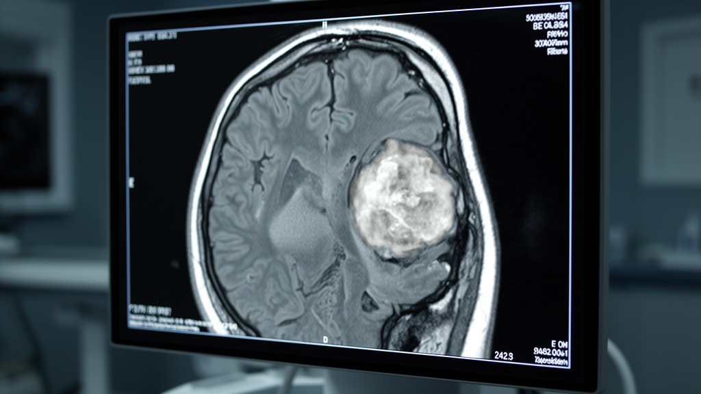
Researchers from India have reported that apparent diffusion coefficient values measured on routine magnetic resonance imaging correlate with histological grade in gliomas and cellularity in meningiomas. The prospective study, published in the Egyptian Journal of Radiology and Nuclear Medicine, analyzed 61 patients with brain tumors to evaluate how diffusion-weighted imaging parameters align with microscopic tissue characteristics. The findings suggest that while diffusion metrics offer a useful supplementary biomarker for preoperative assessment, they lack the precision required to reliably distinguish high-grade gliomas from metastatic deposits on a case-by-case basis.
The study was designed to address the limitations of conventional MRI sequences, which provide excellent anatomical delineation but struggle to predict tumor cellularity or grade before surgery. Diffusion-weighted imaging measures the random displacement of water molecules in tissue, providing a window into cellular packing and extracellular space. When cells crowd together, the extracellular compartment narrows, causing the apparent diffusion coefficient to drop. This inverse relationship between diffusion and cellularity has been observed in stroke imaging and subsequently extended to tumor characterization, though consensus on clinically useful thresholds has remained elusive.
The cohort consisted of 31 males and 30 females, aged 15 to 65 years, who presented with neurological complaints and intracranial mass lesions. Patients with contraindications to MRI were excluded. Of the 61 participants, 47 underwent surgical resection, allowing for direct histopathological correlation. The tumors were classified according to the 2016 World Health Organization Classification of Tumors of the Central Nervous System. Molecular profiling, including IDH mutation analysis and 1p/19q codeletion testing, was not performed, a limitation the authors acknowledge as establishing a pre-molecular baseline for the findings.
Imaging was performed on a 1.5 T scanner using a standardized protocol that included axial T1-weighted, T2-weighted, and fluid-attenuated inversion recovery sequences. Diffusion-weighted echo-planar imaging was acquired at b-values of 0, 500, and 1000 s/mm². Two radiologists, blinded to histopathological results, placed three to five circular regions of interest within the solid enhancing component of each tumor. The mean of these measurements was recorded as ADCmean, while the single lowest value was designated ADCmin. Peritumoral measurements were obtained from non-enhancing T2/FLAIR-hyperintense regions adjacent to the tumor margin.
Measurement reproducibility was assessed in a subset of 20 cases, with intraobserver and interobserver intraclass correlation coefficients of 0.93 and 0.89, respectively, indicating excellent agreement. Among the 26 gliomas identified, 14 were low-grade and 12 were high-grade. Low-grade gliomas demonstrated significantly higher tumoral ADCmin values than high-grade gliomas, with a mean of 1161.57 ± 330.96 × 10⁻⁶ mm²/s compared to 755.50 ± 170.25 × 10⁻⁶ mm²/s. This difference was statistically significant, with a p-value of 0.0008, confirming that lower diffusion values are associated with higher tumor grade.
Receiver operating characteristic analysis of tumoral ADCmin yielded an area under the curve of 0.87, with a 95% confidence interval of 0.74 to 0.99. The optimal cutoff value determined by Youden’s index was 934 × 10⁻⁶ mm²/s, achieving a sensitivity of 83.3% and a specificity of 85.7% for discriminating between low-grade and high-grade gliomas. However, the authors note that individual values overlapped considerably, with low-grade ADCmean ranging from 871 to 1949 × 10⁻⁶ mm²/s and high-grade values spanning 558 to 1233 × 10⁻⁶ mm²/s. This overlap tempers the standalone diagnostic utility of the metric, suggesting it should be interpreted alongside conventional MRI and molecular pathology.
In the 18 meningiomas studied, a strong inverse correlation was observed between tumoral ADCmean and histopathological cellularity, with a Pearson correlation coefficient of r = −0.67 and a p-value less than 0.01. Cellularity was quantified as the average nuclear count across ten high-power fields. The two atypical meningiomas in the cohort fell toward the lower end of the ADC range, but with only two cases, formal statistical comparison between benign and atypical subtypes was not feasible. The physical basis for this correlation is that tightly packed cells leave less extracellular space for water molecules to diffuse through, resulting in lower ADC values.
Conversely, the study found that diffusion parameters failed to distinguish high-grade gliomas from metastases. Neither tumoral ADCmin nor peritumoral ADCmin showed statistically significant differences between the two groups. Tumoral ADCmin was 755.50 ± 170.25 × 10⁻⁶ mm²/s in high-grade gliomas versus 786.00 ± 159.15 × 10⁻⁶ mm²/s in metastases, with a p-value of 0.6491. Peritumoral ADCmin was also indistinguishable, with values of 1465 ± 210 × 10⁻⁶ mm²/s for high-grade gliomas and 1482 ± 216 × 10⁻⁶ mm²/s for metastases. This negative result contradicts some prior proposals that directional diffusion abnormalities in peritumoral tissue could identify regions of tumor cell infiltration.
Densely cellular tumors, including two primary cerebral lymphomas, one medulloblastoma, and two pineoblastomas, consistently exhibited the lowest ADC values in the cohort. The lymphomas had ADCmean values of 397 and 399 × 10⁻⁶ mm²/s, the lowest figures recorded in the study. The authors emphasize that these observations are descriptive rather than generalizable, given the small sample sizes of n = 1 to 2 per tumor type. These findings illustrate a consistent pattern where dense cellular packing tracks with low ADC values across histologically disparate tumor types.
The study has several limitations, primarily the absence of molecular profiling, which is now mandatory for the integrated diagnosis of diffuse gliomas under the 2021 WHO classification. The metastatic subgroup was diagnosed on clinical and imaging grounds without tissue confirmation, introducing a risk of misclassification. The use of a 1.5 T scanner limits signal-to-noise ratio and spatial resolution compared to 3 T platforms, and the specific ADC cutoff of 934 × 10⁻⁶ mm²/s should be considered protocol-specific. The small circular region of interest approach does not capture the full spatial heterogeneity of high-grade neoplasms, and the single-center design limits external generalizability.
Despite these limitations, the prospective design, standardized ROI placement, and blinded image interpretation lend credibility to the principal findings. The data reinforce the position of diffusion-derived ADC as a useful supplementary biomarker that adds value within a multiparametric imaging protocol. The authors suggest that advanced diffusion techniques, such as diffusion tensor imaging and diffusion kurtosis imaging, may overcome some of the limitations of scalar ADC-based approaches and warrant investigation in molecularly characterized cohorts.
In conclusion, ADC measurements from routine diffusion-weighted imaging correlate with glioma grade and meningioma cellularity at the group level. However, considerable inter-group overlap and the inability to separate high-grade gliomas from metastases highlight the need for caution in clinical application. The findings establish a pre-molecular baseline and underscore the importance of integrating diffusion metrics with conventional MRI, molecular pathology, and direct tissue diagnosis for accurate preoperative assessment.
Subject of Research: Correlation of MRI diffusion metrics with brain tumor histology and grade
Article Title: Correlation of apparent diffusion coefficient values with tumor cellularity and grade in brain neoplasms: a prospective study
Article References: Monis, M., Kumar, A., Hasan Arif, S., Huda, M. F., & Khalid, M. (2026). Correlation of apparent diffusion coefficient values with tumor cellularity and grade in brain neoplasms: a prospective study. Egyptian Journal of Radiology and Nuclear Medicine, 57(1), Article 207. https://doi.org/10.1186/s43055-026-01869-y
Image Credits: AI Generated
DOI: 10.1186/s43055-026-01869-y
Keywords: brain tumors, MRI, diffusion-weighted imaging, apparent diffusion coefficient, glioma, meningioma, cellularity, histopathology, neuroimaging, prospective study, Correlation, apparent
Cite Scienmag News
Cassandra Pierce. (November 8, 2026). Brain Tumor MRI Diffusion Values Track Grade and Cellularity. Scienmag. https://scienmag.com/brain-tumor-mri-diffusion-values-track-grade-and-cellularity/
Cassandra Pierce. "Brain Tumor MRI Diffusion Values Track Grade and Cellularity." Scienmag, 8 November 2026, https://scienmag.com/brain-tumor-mri-diffusion-values-track-grade-and-cellularity/. Accessed 8 November 2026.
Cassandra Pierce. "Brain Tumor MRI Diffusion Values Track Grade and Cellularity." Scienmag. November 8, 2026. https://scienmag.com/brain-tumor-mri-diffusion-values-track-grade-and-cellularity/




