


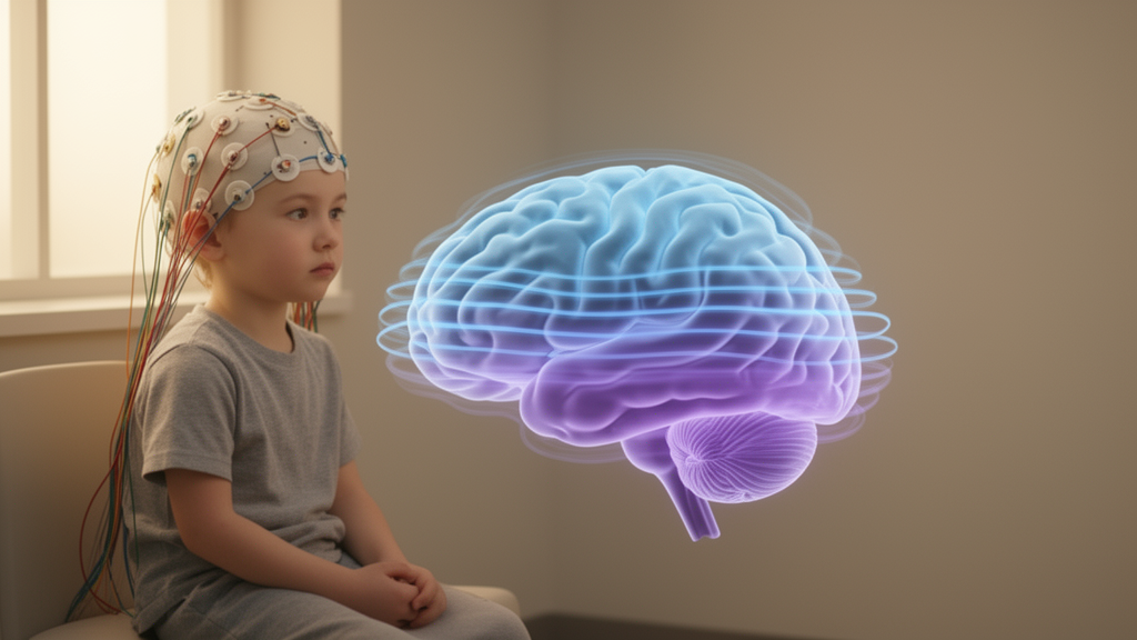
Children with newly diagnosed, untreated childhood absence epilepsy show a subtly different organization of the brain’s alpha rhythm compared with healthy peers, according to a study published in Frontiers in Neuroinformatics. Researchers led by Miroslaw Latka of Wrocław University of Science and Technology analyzed resting, eyes-closed electroencephalogram (EEG) recordings from 49 drug-naive children with childhood absence epilepsy (CAE) and 83 controls, using 19 scalp channels and 26 seconds of data per participant. The findings point to both a slower dominant alpha frequency and a disruption of the normal posterior predominance of alpha-band synchronization in the disorder.
The team focused on two features of the resting EEG. The first was the individual alpha frequency (IAF), the peak frequency of a person’s alpha rhythm, which they estimated using spectral parameterization, a technique that separates the periodic oscillatory components of the power spectrum from the underlying aperiodic background. The second was regional phase-lagged synchronization, measured with the weighted phase-lag index (wPLI), a connectivity metric designed to suppress volume conduction, the artifact by which activity from a single source leaks into multiple electrodes and inflates apparent coupling.
To summarize regional organization, the researchers computed a frontal-to-posterior log-ratio of wPLI values, denoted λFP. In the control children, alpha-band phase synchronization was strongest over posterior regions. To guard against spurious results, they built participant-specific baselines using circular shifting of wavelet coefficients, a surrogate method that destroys genuine temporal relationships while preserving other statistical properties of the signal. Group contrasts were then evaluated both at a fixed frequency of 12 Hz and at each individual’s own alpha peak, among the 131 participants for whom an IAF estimate was available.
The results showed a clear slowing of the alpha rhythm in CAE. The mean individual alpha frequency was 8.80 Hz in patients versus 9.42 Hz in controls, a difference that reached statistical significance at p = 0.002. Because the comparison was confined to drug-naive children, the alteration cannot be attributed to anti-epileptic medication, a common confound in epilepsy EEG studies.
The regional findings were equally striking. In controls at 12 Hz, the posterior predominance of phase-lagged synchronization was pronounced, with a mean λFP of −0.352. After circular-shift baseline subtraction, this value collapsed to −0.036, indicating that roughly 90 percent of the observed contrast reflected genuine frequency-specific organization rather than methodological artifact. In children with CAE, the mean frontal-posterior balance remained near zero, and the observed-minus-surrogate group difference was +0.332 (p < 0.001, effect size g = +0.79), demonstrating a robust loss of the normal posterior alpha focus.
Frequency choice mattered considerably. When the same 131 participants were compared at the fixed 12 Hz band, the group difference after baseline subtraction was +0.316 (p < 0.001). But when each participant was assessed at their own individual alpha frequency, the difference shrank to +0.124 and was no longer statistically significant (p = 0.139). The direct difference between the two contrasts was +0.192 (95 percent confidence interval [+0.017, +0.367], unadjusted p = 0.032), suggesting that part of the apparent CAE-related alteration is tied to the fixed 12 Hz frequency rather than to each child’s true alpha peak.
Global synchronization told a more cautious story. When wPLI was averaged across all electrode pairs rather than contrasted frontally and posteriorly, the group difference after baseline subtraction was −0.021, with a 95 percent confidence interval spanning zero (−0.044 to +0.001, p = 0.069). The authors therefore did not establish a global change in alpha-band phase-lagged synchronization in CAE; the reliable effect was regional in nature.
The study’s conclusions are correspondingly measured. Drug-naive childhood absence epilepsy was associated with slower alpha activity and reduced posterior predominance at 12 Hz, but the regional contrast was smaller at the individual alpha frequency, where a group difference was not established. As a cross-sectional comparison of resting EEG in a single sample, the work characterizes an interictal electrophysiological signature rather than demonstrating a causal mechanism or clinical utility. The modest recording duration of 26 seconds per participant and the fixed-frequency dependence of the effect are limitations the authors themselves highlight through their surrogate-based sensitivity analysis.
Nevertheless, the methodological rigor of the study is notable. By combining spectral parameterization, a volume-conduction-resistant connectivity index, and participant-specific surrogate baselines, the analysis separates genuine frequency-specific network organization from artifacts that can arise in scalp EEG connectivity measurements. The combination of slowed individual alpha frequency and disrupted posterior alpha organization at 12 Hz contributes to a fuller electrophysiological characterization of childhood absence epilepsy, a syndrome in which the interictal, seizure-free brain state appears to carry measurable signatures of the disorder.
Subject of Research: Altered interictal alpha-band EEG organization in drug-naive childhood absence epilepsy
Article Title: Altered interictal alpha-band organization in childhood absence epilepsy
Article References: Latka, M., Glaba, P., Kuryło, M., Jędrzejczyk-Góral, B., Walas, W., Kroczka, S., & Paprocka, J. (2026). Altered interictal alpha-band organization in childhood absence epilepsy. Frontiers in Neuroinformatics, 20, Article 1875377. https://doi.org/10.3389/fninf.2026.1875377
Image Credits: AI Generated
DOI: 10.3389/fninf.2026.1875377
Keywords: childhood absence epilepsy, EEG, alpha rhythm, individual alpha frequency, weighted phase-lag index, phase-lagged synchronization, surrogate analysis, spectral parameterization, electroencephalography, pediatric neurology, brain connectivity, Altered
Cite Scienmag News
Cassandra Pierce. (October 23, 2026). Brain Rhythm Study Finds Slower Alpha Waves in Children With Absence Epilepsy. Scienmag. https://scienmag.com/brain-rhythm-study-finds-slower-alpha-waves-in-children-with-absence-epilepsy/
Cassandra Pierce. "Brain Rhythm Study Finds Slower Alpha Waves in Children With Absence Epilepsy." Scienmag, 23 October 2026, https://scienmag.com/brain-rhythm-study-finds-slower-alpha-waves-in-children-with-absence-epilepsy/. Accessed 23 October 2026.
Cassandra Pierce. "Brain Rhythm Study Finds Slower Alpha Waves in Children With Absence Epilepsy." Scienmag. October 23, 2026. https://scienmag.com/brain-rhythm-study-finds-slower-alpha-waves-in-children-with-absence-epilepsy/


