


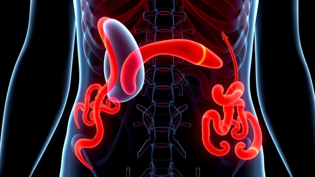
A compression of the artery that feeds the liver, stomach, and spleen has long been one of the most contested diagnoses in pediatric medicine, and a new imaging study suggests that the shape of a child’s body itself may hold part of the answer. Median arcuate ligament syndrome, or MALS, occurs when a fibrous band of the diaphragm presses down on the celiac artery as it emerges from the abdominal aorta, sometimes producing severe stomach pain, weight loss, and other symptoms that baffle clinicians. Now, researchers at Tongji Hospital in Wuhan, China, have used contrast-enhanced computed tomography to show that children and adolescents with suspected MALS differ from their peers not only in the geometry of their arteries and ligaments, but in more fundamental measures of body proportion, including the relationship between abdominal girth and the height of a single lumbar vertebra.
The study, published in BMC Medical Imaging, took the form of a retrospective case-control analysis. The team assembled 45 children and adolescents with suspected MALS and matched them with 45 controls of similar age and sex, all of whom had undergone contrast-enhanced CT scans between January 2015 and July 2023. Rather than relying on a single measurement, the investigators cast a wide net, capturing abdominal circumference, the dimensions of the first lumbar vertebral body, exploratory ratios linking these skeletal landmarks to overall body size, the angle between the abdominal aorta and the celiac artery, and the distances separating the celiac artery and the superior mesenteric artery from the median arcuate ligament itself.
The technical logic behind these measurements is rooted in the anatomy of the upper abdomen. The celiac artery normally branches forward from the aorta at a fairly steep angle, and the median arcuate ligament, a band of fibers that connects the two crura of the diaphragm, typically arcs over the artery without squeezing it. In MALS, that ligament sits lower or the artery runs higher, and the resulting compression can narrow the vessel’s opening. Radiologists have long debated which imaging features truly distinguish pathological compression from harmless anatomical variation, particularly in children, whose bodies are constantly changing as they grow.
What the Chinese team found was striking. Compared with controls, the suspected MALS group had a significantly lower ratio of abdominal circumference to first lumbar vertebral body height, meaning their abdomens were relatively small for their skeletal frame. They also showed a larger angle between the abdominal aorta and the celiac artery, indicating that the artery was deflected more sharply from its parent vessel, and shorter distances between the celiac artery and the median arcuate ligament as well as between the superior mesenteric artery and the ligament. Each of these differences points toward a common theme: in children with suspected MALS, the space available for the celiac artery is tighter, and the vessel itself is bent more dramatically as it exits the aorta.
Crucially, these associations held up after the researchers statistically adjusted for age and sex, two factors that obviously influence body size and vascular anatomy in growing children. A lower abdominal circumference to vertebral height ratio, a larger aorta-celiac angle, and a shorter distance between the superior mesenteric artery and the ligament each remained independently linked to membership in the suspected MALS group. When the investigators combined these variables into a predictive model, it achieved an apparent area under the curve of 0.899 in their development cohort, with a 95 percent confidence interval running from 0.837 to 0.962. In practical terms, a value approaching 0.9 suggests the model can discriminate between suspected MALS patients and controls with high accuracy, though the authors are careful to note that this figure reflects performance on the same data used to build the model and requires external validation.
Perhaps the most intriguing finding concerns age. Within the suspected MALS group, older patients showed greater narrowing of the celiac artery and a larger aorta-celiac angle than younger ones. When the team stratified patients into those aged twelve or younger and those older than twelve, only age emerged as an independent predictor of a higher stenosis category in ordinal logistic regression. Stenosis severity, in turn, correlated positively with age, with vertebral dimensions, and with the aorta-celiac angle, and negatively with the abdominal circumference to vertebral height ratio. In other words, as children grow and their skeletons elongate, the compression of the celiac artery appears to worsen, at least among those already suspected of having the syndrome.
This age-related pattern offers a possible explanation for the clinical course of MALS in adolescents. The condition is most often diagnosed in young, thin patients, frequently teenage girls, and symptoms often prompt imaging during periods of rapid growth. If vertebral growth outpaces the expansion of the abdominal cavity and the ligament-to-vessel spacing fails to widen accordingly, the mechanical squeeze on the celiac artery could intensify during adolescence. The new data lend quantitative support to this idea, suggesting that the interplay between skeletal maturation and vascular compression is not static but evolves through the growing years.
Not every measurement told the same story. The levels at which the visceral arteries originated from the aorta, and the thicknesses of the median arcuate ligament and the diaphragmatic crura, did not differ significantly between the suspected MALS group and the controls. This negative result is informative in its own right, because it suggests that the diagnostic signal lies not in how thick the compressing ligament is or where the arteries branch, but in the spatial relationships between vessels and ligaments and in the overall proportions of the trunk. It also reinforces the idea that MALS is best understood as a problem of geometry rather than of any single structure.
The study’s authors, led by Yufan Wang and corresponding author Yaqi Shen of the Department of Radiology at Tongji Hospital, Tongji Medical College, Huazhong University of Science and Technology, are careful about the limits of their work. This was a retrospective, exploratory analysis of patients with suspected, not surgically confirmed, MALS, and the controls were matched only for age and sex rather than for body size. The apparent performance of the predictive model may be optimistic because it was evaluated within the same cohort used to derive it. The researchers themselves state that independent validation is needed to establish the clinical relevance of their findings, and they frame the measured parameters as complementary information for CT assessment rather than as stand-alone diagnostic criteria.
Even with those caveats, the work adds a fresh dimension to a debate that has often been driven by anecdote and small surgical series. By grounding the discussion in reproducible CT measurements, linking body proportions to vascular compression, and highlighting age as a key modifier of stenosis severity, the study gives radiologists and pediatricians a more structured framework for evaluating children with unexplained abdominal pain. If future prospective studies confirm these associations, the humble ratio of a child’s waistline to the height of a vertebra could become a routine part of the radiology report, helping clinicians decide which young patients truly need further evaluation for this elusive syndrome and which can be reassured that their anatomy, however unusual, is unlikely to be the source of their suffering.
Subject of Research: CT imaging features of median arcuate ligament syndrome in children and adolescents
Article Title: Body proportions and age-related imaging features in children and adolescents with suspected median arcuate ligament syndrome: a CT case-control study
Article References: Body proportions and age-related imaging features in children and adolescents with suspected median arcuate ligament syndrome: a CT case-control study. (n.d.). https://doi.org/10.1186/s12880-026-02826-x
Image Credits: AI Generated
DOI: 10.1186/s12880-026-02826-x
Keywords: median arcuate ligament syndrome, pediatric radiology, celiac artery stenosis, computed tomography angiography, body proportions, abdominal aorta, median arcuate ligament, superior mesenteric artery, case-control study, adolescent abdominal pain, vertebral dimensions, diagnostic imaging
Cite Scienmag News
Ophelia Keating. (September 22, 2026). Body Shape and Age Shape Celiac Artery Compression Clues in Children, CT Study Finds. Scienmag. https://scienmag.com/body-shape-and-age-shape-celiac-artery-compression-clues-in-children-ct-study-finds/
Ophelia Keating. "Body Shape and Age Shape Celiac Artery Compression Clues in Children, CT Study Finds." Scienmag, 22 September 2026, https://scienmag.com/body-shape-and-age-shape-celiac-artery-compression-clues-in-children-ct-study-finds/. Accessed 4 October 2026.
Ophelia Keating. "Body Shape and Age Shape Celiac Artery Compression Clues in Children, CT Study Finds." Scienmag. September 22, 2026. https://scienmag.com/body-shape-and-age-shape-celiac-artery-compression-clues-in-children-ct-study-finds/


