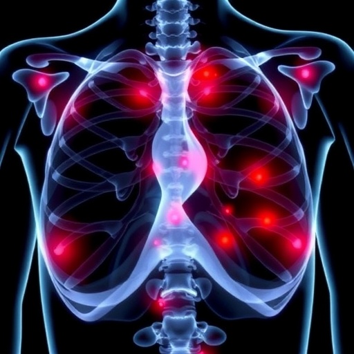
In an inspiring leap forward for cancer research, a recent study unravels the intricate molecular choreography that enables breast cancer cells to colonize bone tissue, opening promising new avenues for therapeutic intervention. This breakthrough centers on the STING-IL6/STAT3 signaling axis, a pivotal pathway sustaining the formation of an osteoclastic niche that facilitates breast cancer bone metastasis. Published in the journal Cell Death Discovery, this pioneering research illuminates the crosstalk between cancer cells and the bone microenvironment, revealing potential targets to disrupt these deadly metastatic processes and improve patient outcomes.
Breast cancer frequently metastasizes to bone, where it causes ulcers of pain, fractures, and profound functional impairment, posing formidable challenges in clinical management. While treatments have evolved, there remains an urgent need for strategies that precisely target the cellular networks enabling tumor cells to infiltrate and remodel bone tissue. This study orchestrates a detailed inquiry into how tumor-derived signals manipulate bone-resorbing cells—osteoclasts—encouraging an environment conducive to cancer growth and survival.
Central to the study is the STING (Stimulator of Interferon Genes) pathway, traditionally known for its role in innate immune sensing of cytosolic DNA and antiviral responses. However, emerging evidence reveals its broader implications in cancer biology and inflammatory signaling. Here, the researchers demonstrate that activation of the STING pathway within breast cancer cells leads to subsequent upregulation of the inflammatory cytokine IL-6, which in turn activates STAT3, a transcription factor implicated in promoting tumor progression and metastasis.
Dissecting this cascade, the study delves into the molecular mechanisms by which STING activation triggers IL-6 secretion, fueling persistent STAT3 phosphorylation in surrounding cells within the bone microenvironment. This signaling axis ignites a vicious cycle that drives osteoclast differentiation and function, catalyzing bone degradation and sculpting an osteoclastic niche where disseminated tumor cells thrive.
The research utilized cutting-edge in vitro and in vivo models to validate these findings, employing breast cancer cell lines, patient-derived xenografts, and genetically engineered mouse models to faithfully recapitulate bone metastasis processes. The team employed molecular inhibition techniques targeting STING as well as IL-6 and STAT3 pathways, observing significant reductions in osteoclast activity and the establishment of metastatic lesions.
One of the most compelling findings relates to the therapeutic potential of small molecule inhibitors and antibodies that disrupt this signaling axis. By selectively blocking components of the STING-IL6/STAT3 pathway, the researchers effectively curtailed the formation of the osteoclastic niche, impeding the invasive capacity of cancer cells in bone and halting metastatic progression. This suggests that a combinatorial approach targeting both tumor-intrinsic pathways and microenvironmental factors could redefine clinical strategies for managing breast cancer bone metastases.
Beyond the molecular insights, the study underscores the complexity of tumor microenvironment interactions, emphasizing how cancer cells hijack physiological pathways like bone remodeling to create protective niches. This paradigm highlights the importance of considering both tumor cells and their surrounding environment in the design of anti-metastatic therapies.
Moreover, the elucidation of STING’s role in this context challenges previous assumptions that it solely exerts anti-tumor effects through immune activation. Instead, this research sheds light on STING as a double-edged sword within tumor biology, capable of promoting a pro-metastatic milieu under specific conditions. This nuanced understanding of STING signaling may spur further investigations into context-dependent modulation of this pathway in different cancer types.
The findings also highlight IL-6 as a critical mediator linking tumor-intrinsic stress responses to systemic inflammatory signaling. Given IL-6’s established role in cancer-related inflammation, these results add depth to our comprehension of how chronic inflammatory circuits foster metastatic niches, reinforcing the notion that inflammatory cytokines serve as promising therapeutic targets.
Pharmacologically, the potential to intercept STAT3 phosphorylation presents a tantalizing therapeutic axis. STAT3, often constitutively activated in various cancers, is notoriously challenging to target due to its intracellular location and pleiotropic functions. Nevertheless, advances in drug design and understanding of STAT3’s regulation may soon allow for precise disruption of its oncogenic activities, obviating deleterious effects on normal tissues.
Insightfully, the work connects the dots between innate immune sensing, chronic inflammation, and bone metastasis, delivering a comprehensive mechanistic framework that integrates previously disparate fields. This holistic approach may inspire the development of multi-targeted therapies capable of dismantling the complex metastatic machinery encoded within tumor and stromal compartments.
Crucially, breast cancer patients suffering from bone metastases endure significant morbidity, and current therapies chiefly focus on symptom management and slowing bone degradation rather than eliminating metastatic clones. Therefore, strategies born from this research hold promise not just for impeding metastatic growth but potentially reversing established lesions through microenvironment modulation.
Future investigations stemming from this work might explore combinatory treatments pairing STING or IL-6/STAT3 pathway inhibitors with conventional chemotherapies, immune checkpoint blockade, or bone-targeting agents such as bisphosphonates and RANKL inhibitors. Such integrated regimens could amplify therapeutic efficacy while mitigating adverse effects by precisely tuning the tumor-bone microcosm.
Additionally, elucidating biomarkers reflecting activation status of the STING-IL6/STAT3 axis in patient-derived samples would enable stratification of patients likely to benefit from targeted therapies. This precision medicine approach could optimize clinical trial designs and expedite translation from bench to bedside.
As metastasis remains the predominant cause of cancer-related mortality, insights into the molecular underpinnings of niche formation empower researchers and clinicians alike to envision treatment paradigms that disrupt cancer’s lethal footholds. This study stands out by marrying immunology and bone biology to tackle a stubborn clinical challenge head-on.
Beyond breast cancer, the implications of STING-IL6/STAT3 signaling in metastatic bone disease potentially extend to other malignancies exhibiting tropism for skeletal tissue, such as prostate and lung cancers. Cross-cancer comparative studies may reveal conserved or unique aspects of these pathways, broadening the impact of this research.
In sum, this groundbreaking research from Zhao, Liu, Kong, and colleagues heralds a new epoch in understanding and combating breast cancer bone metastasis. By clarifying the role of the STING-IL6/STAT3 axis in osteoclastic niche formation, it highlights novel molecular targets with translational potential. As the field advances, integrating these findings into clinical frameworks offers hope for improved survival and quality of life for patients grappling with metastatic breast cancer.
Subject of Research: Breast cancer bone metastasis and molecular signaling pathways involved in osteoclastic niche formation.
Article Title: Therapeutic targeting of STING-IL6/STAT3 axis to inhibit osteoclastic niche formation and breast cancer bone metastasis.
Article References:
Zhao, C., Liu, P., Kong, K. et al. Therapeutic targeting of STING-IL6/STAT3 axis to inhibit osteoclastic niche formation and breast cancer bone metastasis. Cell Death Discov. 11, 483 (2025). https://doi.org/10.1038/s41420-025-02776-3
Image Credits: AI Generated
DOI: https://doi.org/10.1038/s41420-025-02776-3


