Hepatic radioembolization currently unfolds in two separate hospital visits. First comes a pre-treatment procedure in which technetium-99m macro-aggregated albumin (Tc-99m-MAA) is injected into the arteries feeding the liver, so that a scan can reveal how much of the radioactive tracer would escape into the lungs. Only if that lung-shunt fraction (LSF) proves safe, and the particle distribution looks suitable, does the patient return for the actual treatment with yttrium-90 microspheres. A research team now reports that the critical lung measurement can be performed in the intervention room itself, directly after the MAA injection, using a dedicated mobile scanner — a result that could clear the way for merging the two sessions into one.
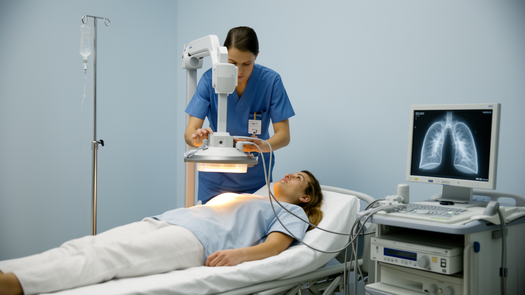
The study, published in EJNMMI Physics, tested a device called IXSI: a mobile dual-layer scanner that combines simultaneous fluoroscopy and nuclear imaging, providing SPECT-like capability alongside cone-beam CT in the angiography suite. Because the instrument is designed for the intervention room, it can acquire planar nuclear images immediately after the Tc-99m-MAA injection, while the patient is still on the table and the interventional team is present. Six patients underwent such direct planar LSF measurements, and for comparison each was also imaged on a conventional SPECT/CT scanner, a Siemens Symbia, in the nuclear medicine department.
The central question was twofold: is such an immediate measurement logistically workable, and is it accurate enough for clinical decisions? Feasibility, the authors report, was achieved — the measurements could be carried out in the intervention room shortly after injection. Accuracy was assessed in two complementary ways. First, digital simulations modeled how the two scanners would perform theoretically, quantifying the expected difference in LSF values between the mobile IXSI system and the conventional SPECT scanner. Second, the actual clinical LSF readings from the six patients were compared between the two modalities.
The simulation results were reassuring. The difference in LSF values between IXSI and the conventional SPECT scanner amounted to −0.3 ± 1.9 percentage points, indicating that, in theory, the mobile intervention-room scanner performs similarly to the established departmental system.
The clinical measurements told a more nuanced story. Uncorrected, the LSF values obtained in the intervention room differed from the conventional SPECT values by 4.5 ± 2.3 percentage points — a gap large enough to matter, since treatment planning hinges on keeping the radiation dose to the lungs within safe limits. The researchers traced this discrepancy not to the scanner hardware but to timing. Radioactive decay and biological redistribution mean that the interval between the Tc-99m-MAA injection and the moment of imaging influences the measured shunt fraction, and the immediate intervention-room scan and the later departmental scan necessarily take place at different times after injection.
To account for this, the team applied a logarithmic correction factor derived from previously published data, adjusting the measurements for the time elapsed between injection and imaging. Once this temporal correction was in place, the difference between the two modalities shrank to 0.0 ± 1.1 percentage points. The clinically observed LSF values thus aligned with what the digital simulations predicted, provided the time difference between injection and imaging was properly corrected for.
The implications reach beyond scanner physics. If the lung-shunt assessment can be completed in the intervention room during the same visit in which the MAA is injected, the traditional two-session structure of hepatic radioembolization — safety evaluation first, treatment later — becomes potentially compressible into a single-session procedure.
The study investigated only six patients, and the published version of this article remains subject to further edits.
Nevertheless, the technical achievement is concrete. A mobile dual-layer scanner bringing simultaneous fluoroscopy and nuclear imaging into the intervention room produced lung-shunt fractions that, after a logarithmic time-interval correction, matched a conventional SPECT/CT scanner to within 1.1 percentage points, while digital simulations had already shown the two systems perform alike. For a therapy whose planning has long depended on a separate nuclear medicine visit, the prospect of obtaining the decisive safety number at the angiography table — minutes after injection — marks a tangible step toward faster, less burdensome radioembolization pathways.
Subject of Research: Measuring the lung-shunt fraction in the intervention room immediately after Tc-99m-MAA injection for hepatic radioembolization planning
Article Title: Lung-shunt fraction measured in the intervention room directly after Tc-99m-MAA injection in hepatic radioembolization
Article References: Dietze, M. M. A., Grosser, O. S., Kerckhaert, C. E. M., Wijnen, N., Smits, M. L. J., Lam, M. G. E. H., Pech, M., & de Jong, H. W. A. M. (2026). Lung-shunt fraction measured in the intervention room directly after Tc-99m-MAA injection in hepatic radioembolization. EJNMMI Physics. https://doi.org/10.1186/s40658-026-00943-8
Image Credits: AI Generated
DOI: 10.1186/s40658-026-00943-8
Keywords: hepatic radioembolization, lung-shunt fraction, Tc-99m-MAA, SPECT, interventional radiology, single-session workflow, nuclear imaging, cone-beam CT, radiation safety, treatment planning, Lung-shunt, fraction
Cite Scienmag News
Denise Maddox. (October 30, 2026). Bedside Lung Shunt Scan Right After MAA Injection Could Enable Single-Session Radioembolization. Scienmag. https://scienmag.com/bedside-lung-shunt-scan-right-after-maa-injection-could-enable-single-session-radioembolization/
Denise Maddox. "Bedside Lung Shunt Scan Right After MAA Injection Could Enable Single-Session Radioembolization." Scienmag, 30 October 2026, https://scienmag.com/bedside-lung-shunt-scan-right-after-maa-injection-could-enable-single-session-radioembolization/. Accessed 30 October 2026.
Denise Maddox. "Bedside Lung Shunt Scan Right After MAA Injection Could Enable Single-Session Radioembolization." Scienmag. October 30, 2026. https://scienmag.com/bedside-lung-shunt-scan-right-after-maa-injection-could-enable-single-session-radioembolization/