In a sweeping breakthrough that redefines our understanding of brain physiology and the blood-brain barrier, a groundbreaking study published in Nature Neuroscience unveils the crucial role of specialized “base barrier cells” in compartmentalizing the choroid plexus, the brain, and the cerebrospinal fluid (CSF). This discovery unfurls a previously uncharted layer of complexity in brain barrier systems, promising revolutionary implications for neurological health, drug delivery, and our fundamental grasp of brain homeostasis.
For decades, the choroid plexus has been recognized as a pivotal interface between the bloodstream and the cerebrospinal fluid, responsible for CSF production and acting as a selective gateway that maintains the brain’s protected environment. However, the mechanisms that precisely maintain this segregation, creating distinct territories within the brain’s anatomy, have remained elusive. This new research illuminates the enigmatic base barrier cells, specialized epithelial cells situated at critical junctures, which act as vital gatekeepers establishing robust compartmental boundaries.
Leveraging an intricate combination of high-resolution imaging, single-cell transcriptomics, and functional assays, the investigative team demarcated the spatial organization and molecular signature of these base barrier cells. The researchers discovered that these cells form a continuous, cohesive epithelial layer strategically located at the base of the choroid plexus. This anatomical positioning allows them to orchestrate the compartmentalization between choroid plexus epithelial structures, the adjacent brain parenchyma, and the cerebrospinal fluid, a function integral to maintaining neural homeostasis and preventing pathological crosstalk.
The molecular architecture of base barrier cells revealed an impressive array of tight junction proteins and signaling molecules that consolidate their barrier function. Notably, these cells express unique combinations of claudins, occludin, and zonula occludens proteins that collectively enhance the selective permeability properties of the base barrier. Moreover, transcriptomic profiling indicated that these cells possess a distinctive gene expression profile that sets them apart from conventional choroid plexus epithelial cells, reflecting an advanced specialization for compartmentalization roles.
Functionally, the study demonstrated that disruption of base barrier cells precipitates profound perturbations in brain-CSF integrity. Experimental ablation or genetic manipulation of these cells led to leakage and mixing of CSF with brain interstitial fluid, underscoring the indispensable role these cells play in preserving cerebrospinal fluid purity. This breach can have cascading effects, potentially triggering neuroinflammation, altered ionic balances, and pathological influxes that could underlie various neurological disorders.
Beyond their barrier function, base barrier cells also appear to engage in bidirectional signaling with immune and neural elements. The researchers uncovered evidence of paracrine signaling molecules released by these cells, which may modulate local immune surveillance and neurovascular dynamics. This revelation opens new avenues for understanding how the brain’s immune environment is tightly regulated at this critical interface, complicating the simplistic view of brain compartments as static zones.
One of the most exciting aspects of this discovery is the potential to leverage base barrier cells as therapeutic targets. Many neurological illnesses, including multiple sclerosis, Alzheimer’s disease, and brain infections, are characterized by disruptions in brain barriers. The newfound knowledge about base barrier cells paves the way for strategies that reinforce, restore, or even selectively bypass these cellular gatekeepers to administer drugs more effectively or mitigate inflammatory damage.
The researchers also posit that the deeper molecular insights into base barrier cells will catalyze advancements in biomimetic barrier models. Traditional in vitro models of the blood-brain barrier have struggled to replicate the full complexity of epithelial interfaces and compartmentalization present in vivo. The identification of this distinct cell type with defined molecular markers and barrier functionalities enables the development of more faithful and predictive platforms for drug screening and neuroscientific exploration.
More broadly, the study challenges the prevailing dichotomous notion of brain-barrier systems as either blood-brain or blood-CSF, introducing a third, refined dimension to our conceptual framework. By highlighting the choroid plexus base barrier cells as a dynamic and functional compartmentalizer, this work calls for a reevaluation of physiological paradigms and fosters a more integrated view of brain fluid dynamics.
From an evolutionary perspective, the presence of these barrier cells might reflect an adaptive innovation for increasingly complex brains, optimizing protection while permitting precise molecular and cellular exchanges. Comparative anatomical studies across species could now seek these cells to understand their conserved roles or species-specific adaptations.
This foundational research also raises compelling questions for future investigation. How exactly do base barrier cells sense and respond to systemic or neural signals? What is their role in aging or neurodegenerative processes? Are there pathological conditions marked by primary defects in these cells? Answers to these questions could open incisive therapeutic windows and predictive biomarkers for brain health.
Furthermore, the study’s multi-disciplinary approach, combining molecular biology, advanced imaging, computational modeling, and physiology, exemplifies the cutting-edge methodologies required to unravel the brain’s labyrinthine architecture. It demonstrates how integrative science can push boundaries to reveal cellular players at scales and in roles previously hidden, setting new standards for brain barrier research.
Critically, this conceptual leap may also inform the development of neuroprotective strategies against environmental toxins, bacteria, and viruses, whose access to the brain is normally tightly regulated. Understanding how base barrier cells enforce compartmentalization may guide interventions in cases such as viral encephalitis or neuroinvasive infections.
In the grand scheme, this revelation marks a pivotal moment in neuroscience, where detailed cellular insights transcend anatomical descriptions to propose new functional templates of brain barrier regulation. It is a call to the scientific community to rethink, reexamine, and reimagine how we define the blood-CSF interface and its guardians, the base barrier cells.
As we anticipate follow-up studies building on this breakthrough, the promise of harnessing base barrier cells to modulate brain environments, enhance drug delivery, and prevent pathological infiltration shines brightly on the horizon. The brain’s elusive compartments have found a new steward, and with it, the horizons of neuroscience research and clinical intervention expand in unprecedented directions.
Subject of Research: Brain barrier systems, choroid plexus, cerebrospinal fluid compartmentalization
Article Title: Base barrier cells provide compartmentalization of choroid plexus, brain and CSF
Article References:
Verhaege, D., De Nolf, C., Van Acker, L. et al. Base barrier cells provide compartmentalization of choroid plexus, brain and CSF. Nat Neurosci (2026). https://doi.org/10.1038/s41593-025-02188-7
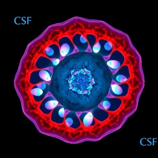
Image Credits: AI Generated
