


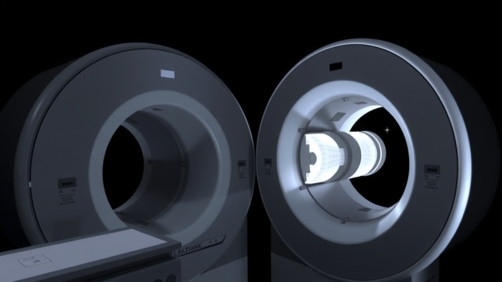
Radiation dose has long been the quiet trade-off behind every cone beam computed tomography scan. Now a team of Finnish researchers reports that a dose-saving technique long considered routine in helical CT can be brought successfully into the world of CBCT, cutting patient exposure by up to 20 percent while actually improving how radiologists judge the resulting images. The study, published in the Annals of Biomedical Engineering, demonstrates for the first time a fully scout-based angular tube current modulation framework for diagnostic cone beam CT, validated on anthropomorphic phantoms and assessed by five board-certified radiologists in blinded paired comparisons.
Cone beam CT has quietly expanded far beyond its dental origins. Orthopedic clinics use it to image extremities, otolaryngologists rely on it for sinus and temporal bone work, and interventional suites employ it for guidance. Yet unlike helical CT, where automatic exposure control and angular tube current modulation, or ATCM, have been standard practice for nearly two decades, most diagnostic CBCT systems deliver a fixed tube current throughout the entire gantry rotation. That means projections passing through thin, low-attenuation regions of the body receive the same radiation as projections that must penetrate dense bone or a broad torso, an imbalance that both wastes dose and degrades image quality where photons run short.
The physics underlying the new method rests on the Poisson statistics of X-ray imaging. Because quantum noise in a projection image scales with the inverse square root of the transmitted photon fluence, the researchers could predict how much current-time product each projection angle would need to hit a uniform target noise level. Rather than relying on the idealized classical model, the team built an empirically calibrated, intensity-dependent noise model using polymethyl methacrylate slabs of varying thicknesses scanned at 80, 100, and 120 kilovolt peaks. The resulting fit captures the nonlinear behavior of a real, polychromatic, energy-integrating detector, including scatter and dark noise, giving the system a reliable bridge between raw detector signal and expected image noise.
To estimate patient attenuation at every angle, the method exploits the low-dose scout views that CBCT scanners already acquire before the main rotation. From posterior-anterior and lateral scout projections, the algorithm extracts median signal intensities within rectangular regions of interest, converts those signals into PMMA-equivalent noise values, and then inverts the calibrated noise model to compute the required current-time product at each scout angle. For intermediate gantry positions between the two scout orientations, an elliptical interpolation model estimates the attenuation profile, an approach long used in CT exposure control that proved remarkably accurate when checked against the fully sampled attenuation measured from all 500 acquired projections.
Because the clinical scanner used in the study, a Planmeca Viso G7, does not yet offer real-time tube current control, the researchers emulated modulation with a projection-library strategy that required no hardware or firmware changes. They acquired a library of CBCT scans of each phantom across a range of exposure levels and, for every projection angle, selected the lowest current-time product that met or exceeded the target modulation profile. Both modulated and unmodulated datasets were then reconstructed identically using a Feldkamp-Davis-Kress algorithm with a Ram-Lak filter and Parker weighting to correct for the scanner’s 210-degree angular coverage, ensuring that any differences in the final images stemmed solely from the distribution of X-ray fluence.
The dose results were striking. With noise magnitude held equal to or lower than that of unmodulated acquisitions, dose-area product fell by 7 to 17 percent across the anthropomorphic phantoms, and Monte Carlo simulations based on the PCXMC platform showed effective dose reductions of up to 20 percent, calculated using ICRP 103 tissue weighting factors with adult and pediatric Cristy-style mathematical phantoms matched to the physical jaw, torso, and thoracic vertebra setups. The mismatch between DAP and effective dose reductions reflects a subtlety of angular dosimetry: organ dose depends on which anatomy lies in the beam path at each angle, so shaving exposure from heavily attenuating views can yield disproportionate effective dose savings.
Just as important as the dose savings was the preservation of image texture. Normalized noise power spectrum analysis of a uniform water phantom showed only negligible differences in both the amplitude and shape of the noise between modulated and unmodulated reconstructions, with no directional noise artifacts appearing in either condition. Noise magnitude, measured as the standard deviation of voxel intensities in Hounsfield units within carefully placed three-dimensional regions of interest, was statistically comparable across protocols. In other words, the technique redistributes photons rather than simply removing them, spending exposure only where the anatomy demands it and banking the rest as dose savings.
The blinded reader study added a human dimension to the quantitative results. Five board-certified radiologists compared 12 matched image pairs spanning low, medium, and high dose levels, each pair pitting a modulated reconstruction against an unmodulated one at fixed noise magnitude, with randomized left-right presentation in a custom web-based viewer. Readers preferred the modulated images in 60 percent of all comparisons, most decisively for torso protocols, where ATCM won 80 percent of paired reads, and jaw protocols, where it won 60 percent. Visually, modulation reduced the lateral streaking and cupping artifacts that typically cluster around dense vertebral bone, improved visibility at vertebral nodes, and sharpened the delineation of thoracic vertebrae in selected protocols. The lone exception was the thoracic vertebra protocol, where readers slightly favored unmodulated images; the authors speculate that unusually smooth reconstructions with sharply defined edges may have been perceived as over-processed.
The authors are careful to frame the work as a proof of concept rather than a finished clinical tool. The library-based emulation does not capture real-world engineering constraints such as tube current slew rate, generator response time, control-loop latency, tube heat loading, and gantry synchronization, and delivering peak exposures at the most attenuating angles could force trade-offs between target image quality, scan time, and motion sensitivity. The elliptical attenuation model may also struggle with asymmetric anatomy or complex pathology, and standardized region-of-interest definition will matter for clinical translation. Still, the framework relies only on raw projection data and existing scout views, making it low-cost and broadly transferable to any CBCT system with raw-data access, and it could combine naturally with optimized scan trajectories, anti-scatter grids, deep-learning scatter correction, and motion-artifact correction algorithms.
The broader implications reach well beyond a single scanner platform. As CBCT pushes into larger fields of view and more radiosensitive anatomical territories, the angular dose imbalances that ATCM corrects grow larger, and the potential benefits grow with them. Scout-based modulation could even reduce the need for patient shielding, which has fallen out of favor in CT partly because it interferes with automatic exposure control. With quantitative noise preserved, effective dose cut by up to a fifth, and radiologists favoring the lower-dose images in most blinded comparisons, the Finnish team has laid out a clear pathway from phantom validation toward hardware implementation and, ultimately, patient-level clinical trials that could make dose-efficient CBCT the norm rather than the exception.
Subject of Research: Angular tube current modulation for radiation dose reduction in diagnostic cone beam computed tomography
Article Title: CBCT Dose Is Reduced with Angular Tube Current Modulation While Maintaining Image Quality and Reducing Photon Starvation Artifacts
Article References: Hyvärinen, T., Onnela, S., Liimatainen, T., Ylisiurua, S., Paakki, J.-J., Järvinen, J., Bode, M. K., Jussila, M.-P., Riekki, V.-P., Hanni, M., & Brix, M. (2026). CBCT Dose Is Reduced with Angular Tube Current Modulation While Maintaining Image Quality and Reducing Photon Starvation Artifacts. Annals of Biomedical Engineering. https://doi.org/10.1007/s10439-026-04354-9
Image Credits: AI Generated
DOI: 10.1007/s10439-026-04354-9
Keywords: angular tube current modulation, cone beam CT, radiation dose reduction, image quality, photon starvation, automatic exposure control, dose-area product, effective dose, noise power spectrum, scout imaging, radiation protection, medical imaging
Cite Scienmag News
Ophelia Keating. (September 12, 2026). Angular Tube Current Modulation Cuts CBCT Radiation Dose Without Sacrificing Image Quality. Scienmag. https://scienmag.com/angular-tube-current-modulation-cuts-cbct-radiation-dose-without-sacrificing-image-quality/
Ophelia Keating. "Angular Tube Current Modulation Cuts CBCT Radiation Dose Without Sacrificing Image Quality." Scienmag, 12 September 2026, https://scienmag.com/angular-tube-current-modulation-cuts-cbct-radiation-dose-without-sacrificing-image-quality/. Accessed 4 October 2026.
Ophelia Keating. "Angular Tube Current Modulation Cuts CBCT Radiation Dose Without Sacrificing Image Quality." Scienmag. September 12, 2026. https://scienmag.com/angular-tube-current-modulation-cuts-cbct-radiation-dose-without-sacrificing-image-quality/


