


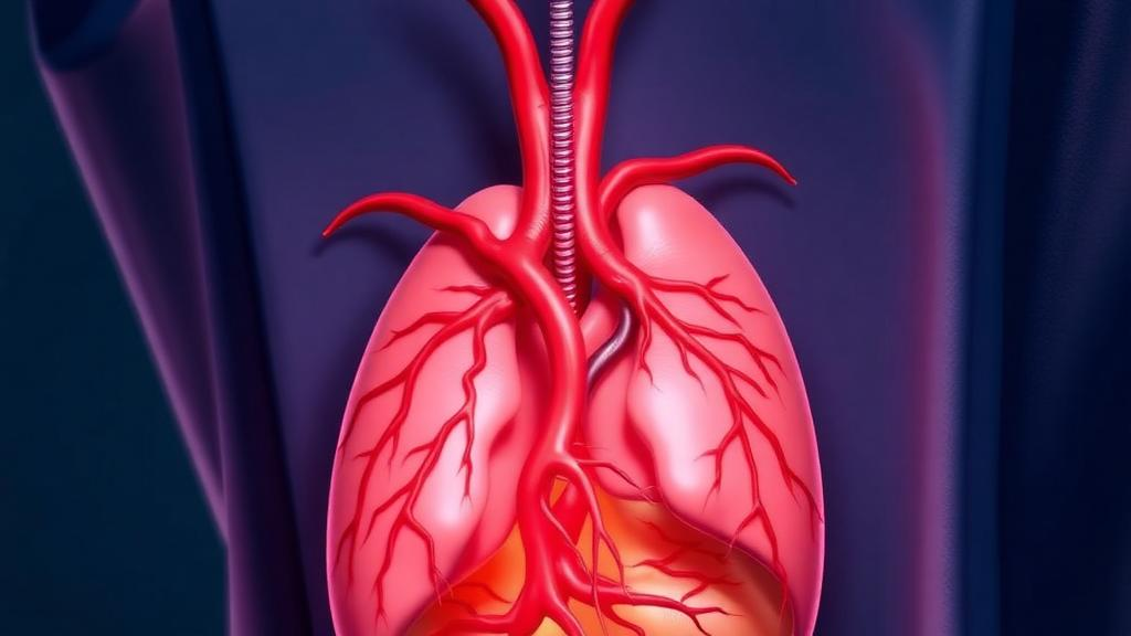
Deep in the chest, a large tumor growing from the thymus gland can do something quietly devastating: it can wrap itself around the superior vena cava, the great vein that carries blood from the head, neck, and arms back to the heart. When that happens, patients develop superior vena cava syndrome, a condition marked by swelling of the face and upper body, headaches, and dangerously impaired venous drainage from the brain. For decades, the standard response was caution. Because the vein itself was involved, many patients were steered toward conservative treatment on the assumption that surgery was too risky to justify. A team at Zhongshan Hospital, Fudan University, in Shanghai, argues that this assumption deserves to be retired, and their new commentary in Clinical Cancer Bulletin lays out, in unusual technical detail, exactly how anesthesia teams can make an operation once considered contraindicated not only feasible but routine enough to have been performed in more than fifty patients.
The operation at the center of the report is called extended resection of thymic tumor combined with artificial bypass between the innominate vein and the right atrial appendage. In essence, surgeons remove the thymic tumor together with the segment of superior vena cava it has invaded, then restore venous outflow by sewing a synthetic vascular graft between the innominate vein and a small pouch-like structure on the heart called the right atrial appendage. Since March 2016, the Shanghai group has explored this approach systematically. A published study from their institution covering fifty-seven patients treated between January 2016 and June 2021 found that the procedure substantially improved quality of life and prolonged survival. Among those cases, twenty-five patients required resection of the left innominate vein alone, two underwent partial resection and repair of the superior vena cava, and thirty received full artificial vessel reconstruction between the innominate vein and the right atrial appendage before the tumor and the vein were removed.
What makes the new commentary valuable is its focus on the anesthetic discipline that underpins the surgery. The authors, anesthesiologists YuanYuan Ma and ShengJin Ge and thoracic surgeon JianYong Ding, emphasize that the operation demands general anesthesia with a single-lumen endotracheal tube, because access is gained through a median sternotomy, the vertical division of the breastbone. Notably, they deliberately avoid combining general anesthesia with an epidural block, a technique often used for pain control in major chest surgery. The reason is practical and unforgiving: these patients require strict anticoagulation around the time of surgery to keep the artificial graft from clotting, and an epidural catheter in a heavily anticoagulated patient carries a risk of spinal bleeding that no surgical benefit can offset.
Preparation begins long before the first incision. The team insists on a mandatory preoperative assessment of both the airway and the great vessels. In their experience, airway integrity is rarely compromised in patients selected for planned surgery, but the vascular assessment is critical: anesthesiologists and surgeons must map precisely where and how extensively the tumor invades the superior vena cava and the brachiocephalic veins, and they must judge whether collateral circulation, the network of smaller veins that develops to bypass a chronically obstructed great vessel, has already been established. The anatomy itself offers a helpful clue for planning. The left innominate vein crosses in front of the aortic arch and follows a long, accessible course, which makes it relatively easy to separate and expose when the tumor has not completely engulfed it. That is why the left innominate vein to right atrium reconstruction is the team’s preferred configuration.
Once the patient is in the operating room, the monitoring strategy reads like a checklist for protecting two vulnerable organs at once: the brain and the heart. The team tracks body temperature and measures the activated clotting time of whole blood, or ACT, at three key moments: before anesthesia induction, fifteen minutes after giving intravenous heparin at a dose of one milligram per kilogram, and thereafter as needed to guide anticoagulation. Depth of anesthesia is monitored with the bispectral index, including the burst suppression ratio, or alternatively with cerebral oxygen saturation, so that any fall in brain perfusion is caught in real time. A catheter is placed retrograde into the cranial segment of the right internal jugular vein to measure venous pressure on the head side of the obstruction, and transesophageal echocardiography is recommended for patients with significant cardiac disease. Vascular access is equally deliberate: the femoral vein and the left radial artery are catheterized, and the right internal jugular vein is cannulated in reverse, toward the head.
The physiological logic that ties the whole anesthetic plan together is captured in a single number. Intracranial perfusion pressure, the pressure driving blood through the brain, is approximately equal to the difference between the mean arterial pressure and the right internal jugular venous pressure. The team therefore watches both values continuously throughout the operation and afterward, and maintains that difference at greater than sixty millimeters of mercury during the entire perioperative period. When the superior vena cava is clamped, venous pressure on the head side can climb steeply, so the team keeps it below thirty millimeters of mercury based on their accumulated experience, elevates the head of the operating table by fifteen to thirty degrees to encourage venous return, and administers intravenous mannitol when appropriate to reduce cerebral swelling. If the bispectral index shows burst suppression appearing or rising, a signal the authors interpret as cerebral hypoperfusion with ischemia, the response is immediate: raise the perfusion pressure, and if necessary change the mode of superior vena cava reconstruction altogether.
Ventilation during the operation follows the principles of lung-protective mechanical ventilation, adapted to the peculiar hemodynamics of venous clamping. The team uses low tidal volumes of four to six milliliters per kilogram of body weight, compensates with an increased respiratory frequency to keep the end-tidal carbon dioxide partial pressure in the normal range, and applies zero positive end-expiratory pressure. That last choice matters: positive pressure in the chest would impede venous return at precisely the moment when the surgical team is rerouting it. Other details reveal how finely tuned the procedure is. During manipulation of the pericardium, the anesthesiologist watches cardiac function, blood pressure, and arrhythmias closely. After the subclavian vein is blocked, any available upper extremity intravenous route is clamped to prevent infusions from being delivered into a territory with no venous outflow. And before the tumor-invaded superior vena cava is clamped, the team confirms that the patient has no peripherally inserted central catheter or subcutaneous port catheter that could be damaged or dislodge.
The moment the bypass graft is opened is one of quiet confirmation. The team observes whether the pressure in the right internal jugular vein drops significantly, which would indicate that the new conduit is successfully draining blood from the head. Heparin is then partially antagonized with protamine, but not fully reversed: the activated clotting time is maintained at an appropriate prolongation, below two hundred seconds, striking a balance between preventing graft thrombosis and avoiding catastrophic bleeding. Throughout the operation the team monitors bleeding, cardiac function, and central venous pressure measured through the femoral venous line, giving blood transfusion, cardiotonic drugs, or diuretics as the situation demands. Cutaneous congestion over the head, neck, and upper chest is treated as a serious warning sign that venous drainage is failing, and the operating surgeon is notified immediately if it appears.
Even the awakening and handover phases carry their own safeguards. As patients emerge from anesthesia, the team watches brain function, including consciousness and movement, and pays attention to respiration, because the right phrenic nerve, which drives one half of the diaphragm, is often transected during the operation, a loss that generally causes little harm but can affect breathing. One instruction the authors say must never be forgotten at the intensive care unit handover is that the right internal jugular vein catheter is for continuous pressure measurement only, never for infusion, since injecting fluid into that vessel would raise intracranial venous pressure. Finally, every patient receives long-term anticoagulation after surgery to keep the artificial graft open, a commitment that begins in the operating room and continues for life.
The larger message of the commentary extends beyond any single operation. Extended resection of thymic tumors with artificial bypass, the authors conclude, requires not only excellent surgical skill but also appropriate intraoperative anesthesia control and intensive critical care, and multidisciplinary teamwork is mandatory. In an era when tumors once deemed inoperable are increasingly being resected with vascular reconstruction, the Shanghai experience offers a template: a standardized anesthetic protocol built around perfusion pressure mathematics, real-time neuromonitoring, meticulous vascular access planning, and disciplined communication between surgeon and anesthesiologist. For patients facing a tumor that has claimed the body’s largest vein, that template may be the difference between palliative care and a genuine chance at extended survival.
Subject of Research: Anesthesia management for extended resection of thymic tumors invading the superior vena cava with artificial innominate vein to right atrial appendage bypass
Article Title: Anesthesia management for extended resection of thymic tumor combined with artificial bypass between the innominate vein and the right atrial appendage
Article References: Ma, Y., Ding, J., & Ge, S. (2024). Anesthesia management for extended resection of thymic tumor combined with artificial bypass between the innominate vein and the right atrial appendage. Clinical Cancer Bulletin, 3(1), Article 14. https://doi.org/10.1007/s44272-024-00020-0
Image Credits: AI Generated
DOI: 10.1007/s44272-024-00020-0
Keywords: thymic tumor, superior vena cava syndrome, anesthesia management, innominate vein bypass, right atrial appendage, vascular reconstruction, thoracic surgery, activated clotting time, bispectral index, cerebral perfusion pressure, anticoagulation, multidisciplinary surgery
Cite Scienmag News
Nathaniel Bowman. (October 4, 2026). Anesthesia Breakthrough Helps Surgeons Rebuild the Superior Vena Cava in Thymic Tumor Surgery. Scienmag. https://scienmag.com/anesthesia-breakthrough-helps-surgeons-rebuild-the-superior-vena-cava-in-thymic-tumor-surgery/
Nathaniel Bowman. "Anesthesia Breakthrough Helps Surgeons Rebuild the Superior Vena Cava in Thymic Tumor Surgery." Scienmag, 4 October 2026, https://scienmag.com/anesthesia-breakthrough-helps-surgeons-rebuild-the-superior-vena-cava-in-thymic-tumor-surgery/. Accessed 4 October 2026.
Nathaniel Bowman. "Anesthesia Breakthrough Helps Surgeons Rebuild the Superior Vena Cava in Thymic Tumor Surgery." Scienmag. October 4, 2026. https://scienmag.com/anesthesia-breakthrough-helps-surgeons-rebuild-the-superior-vena-cava-in-thymic-tumor-surgery/


