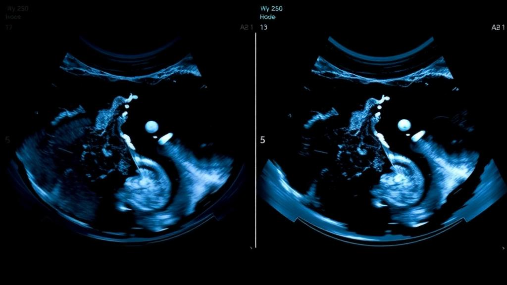
Artificial intelligence has just taken a significant step forward in one of medicine’s most consequential judgment calls: deciding whether a suspicious breast lesion is cancerous or benign. In a prospective multicenter study published in Holistic Integrative Oncology, researchers developed a deep learning system called TGMIL — a temporal-guided, multi-scale, multi-instance learning network — that analyzes entire ultrasound video clips rather than single frozen frames. The results are striking. When the model assisted ultrasonographers of varying experience levels, diagnostic accuracy for difficult BI-RADS category 4 and 5 lesions rose dramatically, with junior sonographers improving from 66.7 percent accuracy to 89.2 percent, and attending-level sonographers climbing from 80.8 percent to 90.0 percent. The study, conducted across four Chinese hospitals, suggests that the future of breast cancer diagnosis may lie not in replacing human experts but in giving them a tireless computational partner that never misses a frame.
The clinical problem the researchers set out to solve is one that radiologists know all too well. Breast lesions classified as BI-RADS 4 or 5 — meaning suspicious enough to warrant biopsy — cover a wide spectrum of malignancy risk, and their ultrasound appearances overlap maddeningly. A benign lesion can look irregular in shape with indistinct margins and a dark, hypoechoic texture; so can a cancer. Posterior acoustic attenuation, a shadowing effect once considered a red flag, appears in both. This morphological ambiguity produces high inter-observer variability, particularly among less experienced practitioners. Ultrasound diagnosis is also highly operator-dependent, shaped by scanning skill, image acquisition standards, and subjective judgment. The stakes are high on both sides: a missed malignancy delays treatment, while an unnecessary core-needle biopsy or surgery exposes a patient to risk, anxiety, and cost for what turns out to be a harmless finding.
The challenge is compounded by breast tissue density. Asian women, on average, have denser breast tissue than other populations, which degrades the performance of mammography, the traditional screening mainstay. In China, clinical guidelines therefore pair mammography with ultrasound as the primary and auxiliary imaging methods for breast cancer detection. Ultrasound is convenient, radiation-free, and particularly effective in dense tissue — but its accuracy in distinguishing benign from malignant BI-RADS 4–5 lesions has remained stubbornly limited. Previous computational efforts largely focused on static images, requiring the sonographer or an algorithm to pick a single representative frame from a live scan. That choice discards a wealth of information: the way a lesion’s boundaries shift as the probe moves, how its internal echoes evolve, and the temporal context that an experienced human eye implicitly absorbs during a real-time examination.
TGMIL was designed specifically to exploit that discarded information. The architecture treats each ultrasound video as a bag of frames in a machine learning framework known as multi-instance learning, where the overall diagnosis is known but the diagnostic value of each individual frame is not. Each frame is first encoded by a VGG-13 convolutional backbone, generating a sequence of frame-level features. Crucially, the model also encodes the temporal order of frames through a linear layer, concatenating this positional information with the visual features so the network can learn from how a lesion appears, evolves, and disappears across the scan. An attention module then adaptively assigns higher weights to the most diagnostically informative frames and aggregates them into a single video-level representation. In effect, TGMIL learns which moments of the scan matter — suppressing redundant frames and emphasizing clinically relevant temporal segments — rather than treating every image equally.
The study enrolled 532 breast lesions categorized as BI-RADS 4–5 across four hospitals between November 2019 and January 2023. Of these, 334 lesions (61 benign and 273 malignant) formed the training and validation sets, while 198 lesions (50 benign and 148 malignant) were reserved as an independent test set. All videos were acquired with high-frequency linear probes, scanned continuously from the first appearance of each lesion to its complete disappearance, and converted to DICOM format. Preprocessing was rigorous: videos were converted to a lossless format, black borders and text overlays were stripped, a K-means algorithm extracted 15 key frames per video, and pixel values were standardized. Every diagnosis was confirmed by pathology from surgery or ultrasound-guided biopsy, and all examinations from the same patient were kept within a single dataset partition to prevent data leakage.
On the external test set, TGMIL achieved an area under the receiver operating characteristic curve of 0.824, decisively outperforming mainstream video classification architectures including 3D-CNN (AUC 0.665), CSN (0.613), and R(2+1)D-50 (0.678). It also beat static image-based classifiers: ResNet50, VGG16, the transformer-based ViT B/32 (which managed only 0.656), and even BU-Mamba, a recently proposed static ultrasound model. BU-Mamba achieved remarkable sensitivity of 0.988 but suffered from low precision of 0.752, indicating a tendency toward over-diagnosis. TGMIL struck a better balance, pairing high sensitivity of 0.966 with precision of 0.836 — meaning it reliably caught malignancies without flooding the clinic with false alarms. Ablation tests confirmed the value of the attention mechanism: simple mean pooling and max pooling baselines reached AUCs of only 0.752 and 0.770 respectively, showing that adaptively identifying informative frames, rather than averaging or maximizing across them, is what drives the performance gain.
The most clinically compelling results came from the reader study. Nine ultrasonographers — three juniors with under three years of experience, three attendings with four to nine years, and three seniors with a decade or more — independently reviewed the 198 test lesions, first without AI assistance and then, four weeks later and in a reshuffled case order, with TGMIL’s real-time benign-versus-malignant probability estimates. Unaided, the model itself achieved a patient-based AUC of 0.845, significantly outperforming both junior sonographers (0.682) and attendings (0.802). With TGMIL’s help, junior accuracy jumped from 66.7 to 89.2 percent and sensitivity surged from 67.4 to 94.2 percent, while attending accuracy rose from 80.8 to 90.0 percent. Across all readers, pooled accuracy improved from 77.5 to 95.0 percent, and the pooled AUC climbed from 0.871 to 0.964. Specificity for the whole group rose from 88.2 to 97.1 percent — a direct strike against unnecessary biopsies.
Interestingly, the most experienced readers gained the least. Senior ultrasonographers, who already achieved an AUC of 0.903 unaided, showed no statistically significant improvement on any metric with AI assistance, and their AUC dipped slightly from 0.898 to 0.885. The authors offer a candid explanation: because TGMIL’s standalone performance (0.824) falls below that of seasoned experts, the model may add little information beyond what veterans already extract from morphology, Doppler signals, and clinical intuition. Worse, when model predictions conflict with expert judgment, uncertain outputs can introduce cognitive interference or automation bias, occasionally nudging a correct decision astray. The study’s failure analysis reinforced this caution: some benign lesions displayed pseudo-malignant features such as spiculated margins that fooled the model into false positives, while some cancers with weak or atypical vascular signals escaped detection as false negatives.
The practical implications are nonetheless considerable. TGMIL was trained on a single NVIDIA RTX 3090 GPU — no supercomputer required — and once trained, inference on ultrasound video is computationally light enough for a local workstation, meaning the system could be integrated directly into the ultrasound workflow. The model also showed strong probability calibration (Brier score 0.071) and robust performance across all four external test centers, with AUCs ranging up to 0.886, suggesting it generalizes across different scanners, hospitals, and patient populations. For regions facing shortages of senior breast imaging specialists, an AI assistant that lifts junior sonographers to near-expert accuracy could meaningfully expand access to reliable diagnosis.
Limitations remain. The cohort was hospital-based and highly selected: only pathologically confirmed BI-RADS 4–5 lesions larger than 5 millimeters were included, excluding prostheses, poor-quality videos, and other BI-RADS categories. No asymptomatic screening population was tested, and screening settings — with lower disease prevalence and smaller incidental lesions — may behave very differently. The authors themselves urge prospective validation in screening cohorts before broader deployment. Still, the study marks a conceptual shift in medical AI: from judging isolated snapshots to understanding the full temporal narrative of a scan, the way a human expert does. If subsequent trials confirm these findings, the humble ultrasound video — long a passive record of a scan — may become an active diagnostic instrument, and the gap between novice and veteran sonographers may finally begin to close.
Subject of Research: Deep learning analysis of ultrasound videos for classifying benign and malignant BI-RADS 4–5 breast lesions
Article Title: AI-enhanced ultrasound video analysis for BI-RADS 4–5 breast lesions: a multicenter study of diagnostic performance
Article References: Li, W.-B., Du, Z.-C., Huang, W.-H., Qiu, R.-Y., Chen, Y.-C., Lu, L.-S., Ji, Z.-Y., Guo, L.-H., He, M.-Q., Wang, L.-S., & Zhang, G.-J. (2026). AI-enhanced ultrasound video analysis for BI-RADS 4–5 breast lesions: a multicenter study of diagnostic performance. Holistic Integrative Oncology, 5(1), Article 80. https://doi.org/10.1007/s44178-026-00302-8
Image Credits: AI Generated
DOI: 10.1007/s44178-026-00302-8
Keywords: artificial intelligence, deep learning, breast cancer, ultrasound, BI-RADS, computer-aided diagnosis, multi-instance learning, medical imaging, diagnostic accuracy, oncology, radiology, multicenter study
Cite Scienmag News
Nathaniel Bowman. (October 11, 2026). AI That Watches the Whole Scan: Deep Learning Reads Breast Ultrasound Videos Better Than Doctors. Scienmag. https://scienmag.com/ai-that-watches-the-whole-scan-deep-learning-reads-breast-ultrasound-videos-better-than-doctors/
Nathaniel Bowman. "AI That Watches the Whole Scan: Deep Learning Reads Breast Ultrasound Videos Better Than Doctors." Scienmag, 11 October 2026, https://scienmag.com/ai-that-watches-the-whole-scan-deep-learning-reads-breast-ultrasound-videos-better-than-doctors/. Accessed 11 October 2026.
Nathaniel Bowman. "AI That Watches the Whole Scan: Deep Learning Reads Breast Ultrasound Videos Better Than Doctors." Scienmag. October 11, 2026. https://scienmag.com/ai-that-watches-the-whole-scan-deep-learning-reads-breast-ultrasound-videos-better-than-doctors/