


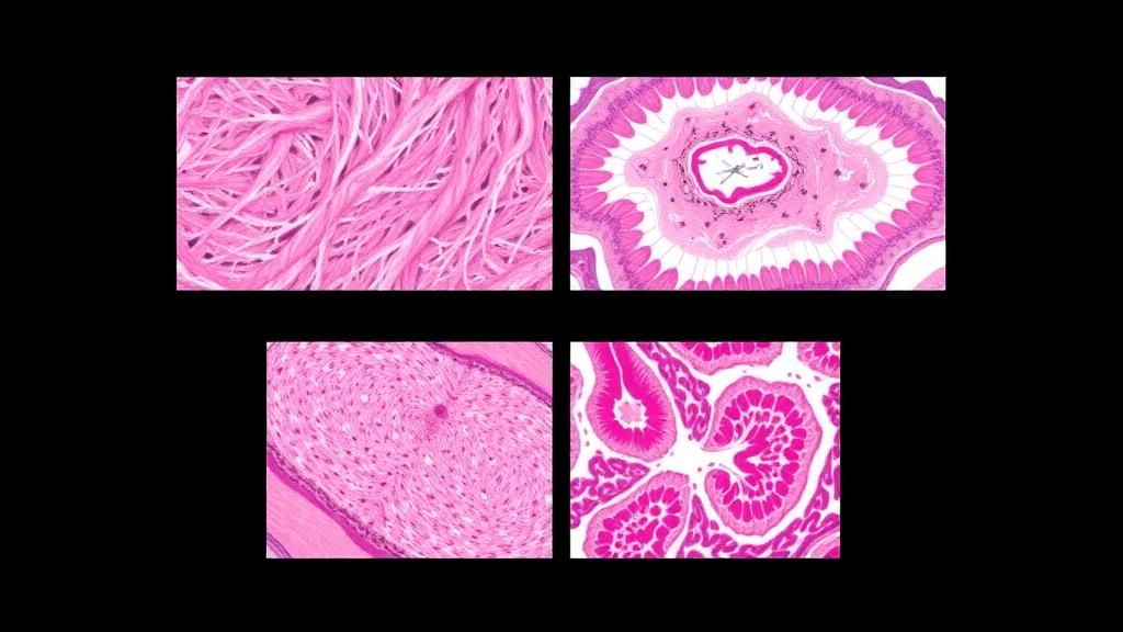
Under a microscope, some of the most troublesome skin diseases look almost indistinguishable. Dermatitis herpetiformis, discoid lupus erythematosus, eczema, and lupus vulgaris all produce inflammatory and granulomatous patterns in skin tissue that can blur into one another even for experienced eyes, and the consequences of confusion are real: each condition demands a different treatment path, from gluten-free diets and dapsone for dermatitis herpetiformis to antitubercular therapy for lupus vulgaris. A new study published in BMC Medical Imaging reports that a deep learning system, built around a well-known neural network architecture, can sort biopsy images of these four conditions with an accuracy of 96.67 percent, while also showing pathologists exactly which parts of the tissue slide drove each decision.
The research, led by Pordil Khan and Abdullah Abdullah of Khyber Medical College in Peshawar, Pakistan, together with collaborators at institutions in Pakistan and Afghanistan, set out to address a gap that has long frustrated dermatopathology, particularly in resource-constrained settings. Machine learning studies on skin lesions have tended to focus on dermatoscopic images of pigmented lesions and skin cancer, where large public datasets exist. Multi-class classification of inflammatory dermatoses from histopathological images, by contrast, has remained comparatively unexplored, even though these conditions are common, diagnostically slippery, and heavily dependent on scarce expert interpretation. The team assembled a balanced dataset of 96 original histopathological images per class, drawn from biopsy specimens collected as part of routine clinical care at Khyber Medical College, with ethics approval from the institution’s review board and written informed consent from patients at the time of biopsy.
Because deep learning models are notoriously data-hungry and 96 images per category is a small foundation, the researchers turned to augmentation, a technique that programmatically expands a training set by applying transformations such as rotations, flips, and other pixel-level adjustments that preserve the diagnostic content of the image. Using the Albumentations library, they applied this process offline and exclusively to the training data, never to the validation images, expanding each class to 486 images. This distinction matters: augmenting only the training set ensures that the models are evaluated on genuine, untouched biopsy images rather than on variations of pictures they have already seen, which would inflate performance estimates and tell clinicians little about real-world behavior.
At the heart of the study was a comparison of four pre-trained convolutional neural networks, each fine-tuned for the four-class task using transfer learning, the strategy of starting from a network already trained on millions of general images and adapting it to a specialized medical problem. ResNet50, a 50-layer architecture whose residual connections allow very deep networks to train stably, emerged as the clear leader, reaching 96.67 percent accuracy on the validation cohort with a micro-average area under the receiver operating characteristic curve of 0.996, a figure indicating near-perfect separation between the disease classes across the full range of decision thresholds. EfficientNet-B0, a compact architecture that scales network depth, width, and image resolution in a balanced way, followed at 95.00 percent accuracy with a micro-average AUROC of 0.990. MobileNetV2, designed for lightweight mobile deployment, managed 85.00 percent, while the older VGG19 architecture trailed at 78.33 percent, a result that reflects how quickly convolutional network design has advanced since VGG’s introduction.
The evaluation protocol deserves attention because it was designed to avoid one of the most common pitfalls in medical image analysis. The models were assessed using an approximately 85:15 patient-level training-validation split, meaning that all images from a single patient were kept on one side of the divide. The validation cohort consisted of 60 original images from 20 patients. Patient-level splitting prevents a model from effectively memorizing the idiosyncrasies of one person’s tissue and then being tested on other slides from the same person, a leakage problem that has produced deceptively high accuracy figures in many published AI studies. Performance was measured with a battery of standard metrics, including accuracy, precision, recall, F1-score, AUROC, the area under the precision-recall curve, and confusion matrices that lay out exactly where the models stumbled.
Those confusion matrices revealed a telling pattern. For ResNet50, every one of the 15 validation images of eczema and every one of the 15 images of lupus vulgaris was classified correctly, while discoid lupus erythematosus and dermatitis herpetiformis each accounted for a single misclassified image among their 15 validation slides. In other words, only two of the 60 validation images were assigned to the wrong disease category, and the errors occurred between conditions that pathologists themselves find difficult to separate. This kind of granular error analysis is far more informative for clinicians than a single headline accuracy number, because it identifies which diagnostic boundaries the algorithm has genuinely mastered and which remain contested territory.
Perhaps the most consequential element of the work, at least for clinical acceptance, is its use of explainability techniques. Deep networks are often criticized as black boxes: they produce a label but no rationale, and a pathologist asked to trust an opaque verdict on a biopsy is likely to decline. The researchers applied Grad-CAM++, a gradient-based visualization method that generates heatmaps highlighting the image regions most responsible for a model’s prediction. When these heatmaps are overlaid on the original histopathological image, they show whether the network is attending to diagnostically meaningful structures, such as the characteristic neutrophilic deposits at the dermal papillae seen in dermatitis herpetiformis or the granulomatous inflammation of lupus vulgaris, or whether it is latching onto irrelevant artifacts like staining variation or cutting marks. Qualitative visualization of this kind turns the model from an oracle into a colleague whose reasoning can be inspected, checked, and, when necessary, overruled.
To move the work beyond a laboratory exercise, the team also built a web-based prototype as an AI-assisted research tool for supportive image classification. Such prototypes are a familiar bridge in the medical AI literature: they demonstrate that a trained model can be packaged into an interface that a clinician or researcher could actually use, uploading a histopathological image and receiving a classification alongside its explanation. The authors are careful, appropriately, to frame this as a research prototype rather than a diagnostic device, and the study’s own limitations section is candid about why. The dataset was small and came from a single center, and there was no external validation on images from other hospitals, scanners, or staining protocols, all of which are known to degrade model performance when conditions differ from the training environment.
Those caveats do not diminish the significance of the demonstration; they define the roadmap. The authors explicitly call for future studies to evaluate larger, multi-center datasets and to incorporate additional clinical information, such as patient demographics and laboratory findings, which could be fused with image features to improve robustness. The distinction between feasibility and clinical applicability is one that the field of medical imaging AI has had to learn repeatedly, and this study draws it honestly. What the results establish is that the diagnostic signal separating these four inflammatory dermatoses is present in histopathological images at a strength that modern convolutional networks can extract, even from a modestly sized, single-institution dataset, provided that training is handled carefully with augmentation, transfer learning, and patient-level validation.
The broader implications reach well beyond dermatopathology. In many parts of the world, expert histopathologists are scarce, and diagnostic delays for conditions like cutaneous tuberculosis or lupus can be measured in months or years. A tool that offers a rapid, explainable second opinion on a biopsy image, flagging the most likely diagnosis and pointing to the tissue regions that support it, could help prioritize cases, reduce errors, and support training of junior pathologists. The finding that ResNet50 and EfficientNet-B0, both freely available architectures, achieved AUROC values above 0.99 on this task suggests that the barrier is not the sophistication of the model but the availability of well-curated, diverse data. If subsequent multi-center studies confirm these results, explainable deep learning could become a routine assistant at the microscope, transforming one of pathology’s most subjective judgment calls into a transparent, quantifiable, and auditable process, and bringing that capability first to the settings where expert eyes are hardest to find.
Subject of Research: Explainable deep learning classification of inflammatory skin diseases from histopathological images
Article Title: Explainable deep learning-based multi-class classification of inflammatory dermatoses from histopathological images
Article References: Khan, P., Abdullah, A., Ahmed, M., Ullah, Q. M. F., Umer, H., Qasim, M., Aizad, G., Gandapur, T. K., & Talha, M. (2026). Explainable deep learning-based multi-class classification of inflammatory dermatoses from histopathological images. BMC Medical Imaging. https://doi.org/10.1186/s12880-026-02897-w
Image Credits: AI Generated
DOI: 10.1186/s12880-026-02897-w
Keywords: deep learning, histopathology, dermatitis herpetiformis, discoid lupus erythematosus, eczema, lupus vulgaris, ResNet50, Grad-CAM++, transfer learning, dermatopathology, medical imaging AI, Pakistan
Cite Scienmag News
Blake Davidson. (October 6, 2026). AI Reads Skin Biopsies: Deep Learning Tells Four Look-Alike Inflammatory Diseases Apart. Scienmag. https://scienmag.com/ai-reads-skin-biopsies-deep-learning-tells-four-look-alike-inflammatory-diseases-apart/
Blake Davidson. "AI Reads Skin Biopsies: Deep Learning Tells Four Look-Alike Inflammatory Diseases Apart." Scienmag, 6 October 2026, https://scienmag.com/ai-reads-skin-biopsies-deep-learning-tells-four-look-alike-inflammatory-diseases-apart/. Accessed 6 October 2026.
Blake Davidson. "AI Reads Skin Biopsies: Deep Learning Tells Four Look-Alike Inflammatory Diseases Apart." Scienmag. October 6, 2026. https://scienmag.com/ai-reads-skin-biopsies-deep-learning-tells-four-look-alike-inflammatory-diseases-apart/


