


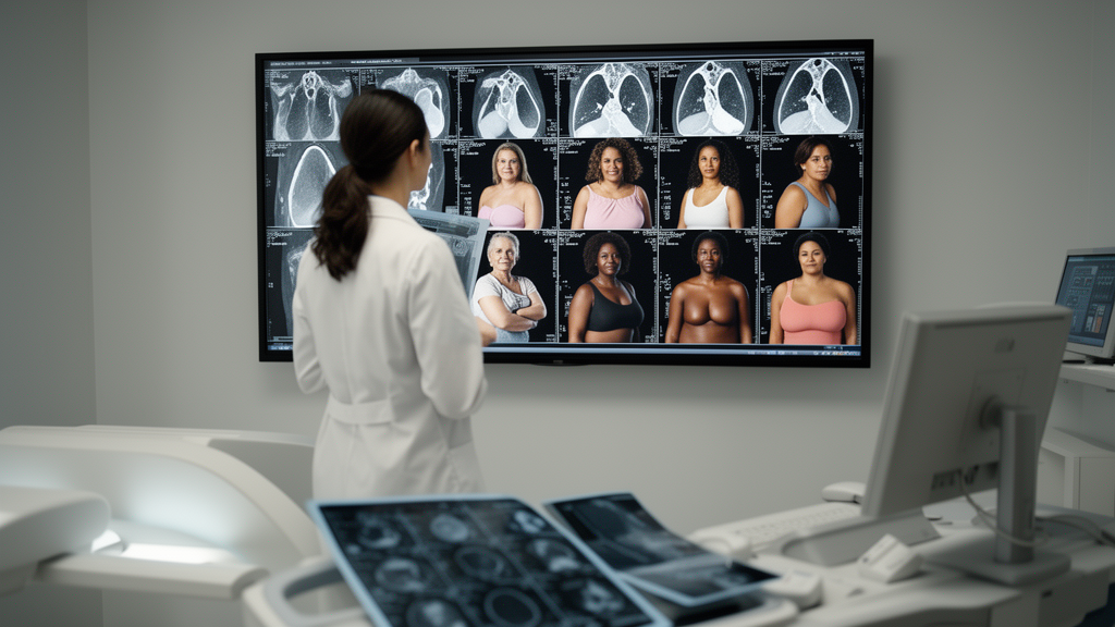
Artificial intelligence systems for detecting breast cancer on mammograms can report accuracy above 95% on benchmark datasets, yet their performance can fall when the same models encounter independent clinical data. A 2026 review concludes that dataset size, annotation quality, demographic representation and geographic coverage—not just increasingly complex neural-network architecture—remain major determinants of whether these tools generalize reliably. The analysis warns that high results on small, geographically concentrated collections should not be mistaken for evidence of broad clinical performance.
The review examines publicly available mammography datasets and their influence on the development of machine-learning and deep-learning systems for classification, lesion detection and segmentation. It covers conventional feature-based methods, convolutional neural networks, transfer learning, object detectors, segmentation networks, Vision Transformers, multimodal systems, explainable AI and privacy-preserving approaches. A structured narrative search covered studies published from 2016 through 2025, with preference given to research using public mammography collections. The authors also assessed each dataset qualitatively for scale, modality, annotation availability and label richness; those suitability ratings were not experimental performance scores.
Public collections have expanded markedly over three decades, but their capabilities remain uneven. MIAS, released in 1994, contains 322 film mammograms from 161 patients with image-level labels and has become a simple benchmark for conventional algorithms and lightweight convolutional networks. DDSM and its curated version CBIS-DDSM provide larger collections with pathology information, lesion regions and, in the curated dataset, segmentation masks. INbreast contains 410 high-quality full-field digital mammograms from 115 patients with pixel-level masks, making it valuable for lesion detection and segmentation research but too small for training large models from scratch. These older resources also rely heavily on digitized film or geographically limited populations.
More recent repositories offer scale, richer labels and, in some cases, clinical context. VinDr-Mammo contains 5,000 Vietnamese full-field digital examinations and 20,000 images with BI-RADS assessments and bounding boxes. CMMD includes 3,712 images from 1,775 Chinese patients, histopathology labels and molecular-subtype information, although it lacks region-of-interest annotations. OMI-DB provides a very large UK collection with lesion annotations, clinical metadata and longitudinal screening information, but access is restricted. DMID, derived from an Indian population, includes 510 full-field digital images with lesion and clinical metadata and remains comparatively limited and evolving. Together, these resources have enabled more ambitious research, but no single public collection combines large scale, broad demographics, standardized annotation, comprehensive clinical metadata and open access.
Annotation depth determines which questions an AI system can learn to answer. Whole-image labels support image classification but provide no clue about where a suspicious finding lies. Bounding boxes identify approximate lesion locations for detectors such as YOLO and Faster R-CNN, while pixel-level masks delineate boundaries for segmentation models such as U-Net and Mask R-CNN. Multi-level annotations can support systems that jointly classify, locate and outline lesions. Standardized BI-RADS descriptors, covering findings such as lesion shape, margins, calcifications and breast density, can add consistent clinical semantics. Yet annotation formats and labeling practices differ across collections, and inter-observer variability introduces label noise. Expert-drawn pixel masks are also labor-intensive, limiting the scale of precisely annotated data.
The technical trajectory has moved from manually engineered texture, shape and statistical features to end-to-end deep networks. Convolutional neural networks learn hierarchical image representations and remain the dominant approach, while transfer learning reuses pretrained weights to cope with smaller labeled collections. YOLO detectors favor speed and real-time inference, whereas Faster R-CNN generally provides stronger localization at greater computational cost. U-Net variants produce detailed lesion boundaries but depend on scarce pixel-level annotations. Vision Transformers use self-attention to capture long-range spatial relationships, and hybrid CNN–Transformer models combine local texture with global context. The review notes, however, that transformers usually require more training data than CNNs. Ensembles and multi-stage systems can integrate several tasks, but their computational complexity may complicate routine clinical use.
High benchmark results do not resolve the central generalization problem. The reviewed literature contains many reports of accuracy above 90%, and some studies describe values above 98% on benchmark collections. Such figures concern the datasets, preprocessing and evaluation protocols used in those studies and do not establish equivalent performance in hospitals with different equipment, acquisition protocols, patient populations or annotation practices. Models evaluated on an independent dataset can perform less well, particularly when breast density, disease distribution, image characteristics or preprocessing differ. Transfer learning, domain adaptation and ensemble methods may reduce these shifts, but the review says they do not eliminate dataset bias, class imbalance and limited interpretability.
Geographic concentration compounds the problem. Much of the research literature relies on North American and European collections, particularly DDSM, CBIS-DDSM, MIAS and INbreast. Public data from Africa, South America and parts of Asia remain scarce, including in India despite its substantial breast-cancer burden. DMID is an important step toward regional representation, but its institutional and geographic coverage is not yet sufficient. Private hospital datasets may contain richer clinical information and greater patient diversity, yet restricted access prevents reproduction and standardized comparison. This imbalance makes it difficult to determine whether a model trained in one population will retain useful sensitivity and specificity in another, or whether domain differences could increase false-positive or false-negative predictions.
The review therefore calls for a comprehensive, geographically diverse mammography benchmark assembled across institutions and healthcare settings. Such a resource would need consistent BI-RADS labels, lesion localization, segmentation where feasible, pathology and follow-up information, and clearly documented data splits and evaluation metrics. Privacy-preserving collaboration through federated learning could allow institutions to contribute to model development without centralizing sensitive patient data. Prospective, multicenter validation would also be needed to test whether benchmark performance survives contact with routine clinical workflows. The central message is that AI-assisted mammography may become more capable through transformers, multimodal learning and explainable systems, but trustworthy deployment depends on data that represent the people and conditions the software is intended to serve.
Subject of Research: How dataset quality, demographic bias and annotation standards affect AI-based mammography for breast cancer detection
Article Title: Mammography datasets and artificial intelligence for breast cancer detection: challenges, bias, and future directions
Article References: Mohideen, A. K., Prakash, P. N. S., & Priyadharshini, M. K. (2026). Mammography datasets and artificial intelligence for breast cancer detection: challenges, bias, and future directions. Frontiers in Artificial Intelligence, 9, Article 1933770. https://doi.org/10.3389/frai.2026.1933770
Image Credits: AI Generated
DOI: 10.3389/frai.2026.1933770
Keywords: mammography, breast cancer, artificial intelligence, medical imaging, dataset bias, deep learning, Vision Transformers, annotation standards, external validation, federated learning, datasets, artificial
Cite Scienmag News
Blake Davidson. (October 20, 2026). AI Mammography Advances Falter When Datasets Lack Diversity. Scienmag. https://scienmag.com/ai-mammography-advances-falter-when-datasets-lack-diversity/
Blake Davidson. "AI Mammography Advances Falter When Datasets Lack Diversity." Scienmag, 20 October 2026, https://scienmag.com/ai-mammography-advances-falter-when-datasets-lack-diversity/. Accessed 20 October 2026.
Blake Davidson. "AI Mammography Advances Falter When Datasets Lack Diversity." Scienmag. October 20, 2026. https://scienmag.com/ai-mammography-advances-falter-when-datasets-lack-diversity/


