


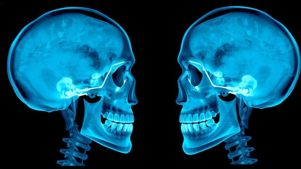
Aligning an X-ray of a patient’s skull with a photograph of that same patient’s face sounds like a problem that should have been solved decades ago. Yet for all the progress in medical imaging and artificial intelligence, the task of precisely overlaying a lateral cephalometric radiograph—the side-view X-ray used in orthodontics for nearly a century—with a facial profile photograph remains stubbornly difficult. A new study published in Medical & Biological Engineering & Computing by researchers at Sichuan University, working across computer science and clinical stomatology, presents a carefully engineered framework that tackles this cross-modality alignment problem and, crucially, proposes a way to measure success that clinicians can actually trust.
The challenge stems from a fundamental mismatch between the two imaging modalities. A cephalometric radiograph captures the bony architecture of the head along with soft tissue shadows, magnified by the geometry of the X-ray beam and acquired with the patient’s head held in a standardized but individually variable position. A facial photograph, by contrast, records only the skin surface, under different lighting, with its own perspective distortions and scale. The two images differ in resolution, contrast, texture and anatomical content, which means the classic image-similarity measures that drive many registration algorithms—such as mutual information, a staple of multimodal medical image alignment since the late 1990s—struggle to find reliable correspondences between them.
The Sichuan University team, led by corresponding author Yuanyuan Chen of the College of Computer Science together with clinical collaborators from the West China Hospital of Stomatology, approached the problem not by building an ever-larger deep learning model, but by combining high-resolution anatomical landmark localization with a mathematically constrained transformation. The framework first detects keypoints—corresponding anatomical features such as recognizable landmarks on the facial profile and their radiographic counterparts—using techniques that can localize these points at high resolution. It then performs a weighted two-dimensional similarity registration based on the Procrustes framework, a classical statistical method for finding the optimal rotation, translation and uniform scaling that best matches one set of points to another.
The deliberate restriction to a similarity transform is a key design decision, and it reflects clinical reasoning rather than computational convenience. X-ray acquisition introduces a global magnification factor that varies from machine to machine and patient to patient, so a uniform scale factor is genuinely needed to accommodate that difference. But shearing and local non-rigid deformation—warps that many modern registration pipelines happily apply—are deliberately excluded. In craniofacial analysis, an algorithm that bends the anatomy to fit would undermine the diagnostic meaning of the overlay. By constraining the transformation to physically plausible motions, the framework ensures that what it aligns is anatomically interpretable.
Interpretability is reinforced by the way the registration is weighted. Rather than treating every detected keypoint as equally reliable, the researchers incorporate expert-defined anatomical priors as weights in the transformation process. Landmarks that clinicians consider more diagnostically important, or that can be localized with greater confidence, exert proportionally more influence over the final alignment. This is a form of knowledge injection that bridges the gap between purely data-driven methods and the accumulated expertise of orthodontic practice, and it echoes a broader trend in medical image analysis where weak supervision and anatomical awareness are increasingly used to keep algorithms honest.
Perhaps the more provocative contribution, however, lies in how the framework is evaluated. The authors argue that the field lacks clinically validated evaluation criteria for this kind of cross-anatomy registration, and they respond by introducing an adaptive seed-driven contour tracking algorithm, abbreviated ASDCT, for precise performance assessment. Instead of relying solely on landmark displacement errors, the evaluation traces the soft-tissue facial profile contour in both images and measures how well the registered contours coincide. Two headline metrics emerge: Contour Distance, and Contour Dilation Overlap, which quantifies how much of one dilated contour region is covered by the other.
The results, obtained on 198 paired clinical samples, are striking for their stability. The framework achieves a Contour Distance of 3.52 pixels and a Contour Dilation Overlap of 87 percent, and, more importantly, these computational metrics correlate strongly with expert clinical assessments. That correlation is the detail that matters most, because the registration literature has long suffered from a validation problem. Researchers have repeatedly shown that widely used surrogate metrics, such as tissue overlap measures borrowed from segmentation, do not reliably track true registration accuracy—a concern raised prominently in the literature for over a decade. By demonstrating that their contour-based measures track what experienced clinicians actually judge to be a good alignment, the team offers a reproducible evaluation standard rather than another leaderboard number.
The clinical payoff of robust cross-modal alignment is the integrated craniofacial analysis that orthodontics has long pursued. Orthodontists routinely reason about the relationship between the facial soft tissue envelope and the underlying skeletal structure: how far the chin sits relative to the jaw, how the lips drape over the teeth, how growth or surgical intervention will reshape the profile. Today those judgments are made by mentally fusing the radiograph and the photograph. A reliable automated overlay turns that mental act into a quantitative one, enabling treatment planning, outcome prediction and communication with patients to be grounded in a single, aligned representation of both bone and surface anatomy.
The study also situates itself intelligently within the current wave of deep learning registration methods. Frameworks such as VoxelMorph demonstrated that convolutional networks can learn deformable alignments from data, and more recent foundation-model approaches promise multimodal registration across diverse acquisition conditions. But the authors’ stance is pragmatic: rather than asking a black-box network to solve the entire alignment, they decompose the problem into reliable keypoint detection, a constrained geometric solution, and expert-informed weighting, with deep learning contributing where it is strongest. The result is a pipeline whose each stage can be inspected, validated and trusted—a design philosophy that may prove as influential as the numbers themselves.
Ethical safeguards and data handling also receive attention. The study was approved by the Ethics Committee of West China Hospital of Stomatology at Sichuan University, written informed consent was obtained from all participants, and the peri-orbital eye regions of facial photographs were masked to de-identify subjects before analysis and publication. In an era when facial images are biometric identifiers, that attention to privacy signals a maturing standard for clinical computer vision research. Taken together, the work suggests that the future of medical image registration may lie less in ever-larger models than in frameworks that marry precise landmark technology, anatomically meaningful constraints, and evaluation metrics that physicians recognize as their own—precisely the bridge between computational precision and clinical practice that this team set out to build.
Subject of Research: Cross-modality registration of cephalometric radiographs and facial profile photographs for craniofacial analysis
Article Title: Robust cross-anatomy image registration leveraging keypoint priors and contour-based assessment
Article References: Xu, Y., Zeng, H., Xue, C., Xu, H., & Chen, Y. (2026). Robust cross-anatomy image registration leveraging keypoint priors and contour-based assessment. Medical & Biological Engineering & Computing. https://doi.org/10.1007/s11517-026-03681-2
Image Credits: AI Generated
DOI: 10.1007/s11517-026-03681-2
Keywords: image registration, cephalometric radiographs, facial profile photographs, craniofacial analysis, keypoint detection, Procrustes analysis, contour tracking, medical image analysis, orthodontics, multimodal alignment, evaluation metrics, artificial intelligence
Cite Scienmag News
Denise Maddox. (October 5, 2026). AI Framework Aligns Skull X-Rays With Facial Photos for Orthodontics. Scienmag. https://scienmag.com/ai-framework-aligns-skull-x-rays-with-facial-photos-for-orthodontics/
Denise Maddox. "AI Framework Aligns Skull X-Rays With Facial Photos for Orthodontics." Scienmag, 5 October 2026, https://scienmag.com/ai-framework-aligns-skull-x-rays-with-facial-photos-for-orthodontics/. Accessed 5 October 2026.
Denise Maddox. "AI Framework Aligns Skull X-Rays With Facial Photos for Orthodontics." Scienmag. October 5, 2026. https://scienmag.com/ai-framework-aligns-skull-x-rays-with-facial-photos-for-orthodontics/


