


Brain tumors are among the most feared diagnoses in medicine, and the scans that reveal them are only as useful as the eyes that read them. Radiologists spend years training to spot the subtle signatures of gliomas, meningiomas, and pituitary tumors on magnetic resonance imaging, yet even experts can disagree, and manual interpretation of thousands of slices is slow and mentally exhausting. A new study from researchers at Manipal Institute of Technology and NMAM Institute of Technology in India, published in Discover Artificial Intelligence, describes a unified deep learning framework that classifies brain tumors, delineates their boundaries, and explains its own reasoning — all within a single automated pipeline.
The team, led by Rashmitha R. Nayak and B. N. Anoop, tackled a problem that has long fragmented the field of medical image analysis. Most previous studies have treated tumor classification and tumor segmentation as separate challenges, and explainability tools have typically been bolted on afterward rather than woven into the diagnostic workflow. The new framework integrates three components into one system: an ensemble classifier that decides whether a scan shows a glioma, meningioma, pituitary tumor, or healthy tissue; a three-dimensional U-Net that traces the exact extent of the tumor; and Grad-CAM visualizations that highlight which parts of the image drove the model’s decision.
The classification engine is built on two well-known convolutional neural network architectures, DenseNet121 and InceptionV4, combined through a technique called soft voting. DenseNet121 uses a dense connectivity pattern in which every layer receives inputs from all preceding layers, encouraging feature reuse and stable gradient flow during training. InceptionV4, a deeper member of the Inception family, balances computational cost and representational power through carefully factorized convolutional blocks and residual connections. Rather than forcing a choice between them, the ensemble averages the probability distributions each model assigns to the four diagnostic categories and selects the class with the highest combined score. The intuition is simple: two networks that see the image differently are less likely to make the same mistake.
The numbers back that intuition up. On a held-out test set drawn from a publicly available Kaggle dataset of 7,023 grayscale MRI images, the ensemble achieved 98.52 percent accuracy, edging out InceptionV4 at 98.28 percent, DenseNet121 at 97.93 percent, and a lightweight custom CNN baseline at 95.00 percent. Per-class precision, recall, and F1-scores were consistently high, with a macro-averaged F1 of roughly 0.98, indicating that the model performed well across all four categories rather than excelling on common classes while neglecting rare ones. Receiver operating characteristic analysis reinforced the picture, with macro and micro AUC values approaching 1.0 for the transfer learning models and the ensemble.
The researchers were careful about statistics, running stratified five-fold cross-validation to tune hyperparameters and then applying a Friedman test to the fold-wise accuracies. That test confirmed a statistically significant difference among the four classifiers, with the ensemble consistently ranked first. Pairwise Wilcoxon signed-rank tests, however, did not reach significance, a limitation the authors attribute to the small number of folds, which constrains the statistical power of non-parametric comparisons even when the ranking is stable. Cross-validation standard deviations of just 0.03 to 0.08 percentage points suggested that both flagship networks behave consistently across data splits.
Classification alone tells a clinician what a tumor probably is; segmentation shows where it is. For that second task, the team trained a 3D U-Net on the BraTS 2020 dataset, a collection of multimodal MRI volumes with expert-annotated tumor masks. Each subject’s scans included T1, T1-contrast, T2, and FLAIR sequences, of which the researchers used the latter three as input channels. The U-Net’s encoder-decoder structure, connected by symmetric skip connections, lets the network capture both high-level context and fine spatial detail, and a final sigmoid layer produces a voxel-wise probability map of the whole tumor region. The authors simplified the problem to binary segmentation by merging the enhancing tumor, tumor core, and whole tumor subregions into a single foreground class.
Training the segmentation network required a loss function tailored to the peculiar challenges of medical imaging, where tumor voxels are vastly outnumbered by healthy tissue. The team combined binary Dice loss and focal loss in equal measure, a hybrid designed to maximize spatial overlap while down-weighting the easy background examples that would otherwise dominate the gradient signal. The resulting model achieved a Dice similarity coefficient of 0.8423 and an Intersection over Union of 0.7497 on the independent test set of 105 MRI volumes. Qualitative inspection showed close agreement with ground-truth annotations for larger, well-defined tumors, while small or diffuse boundaries remained harder, a difficulty familiar from prior BraTS studies.
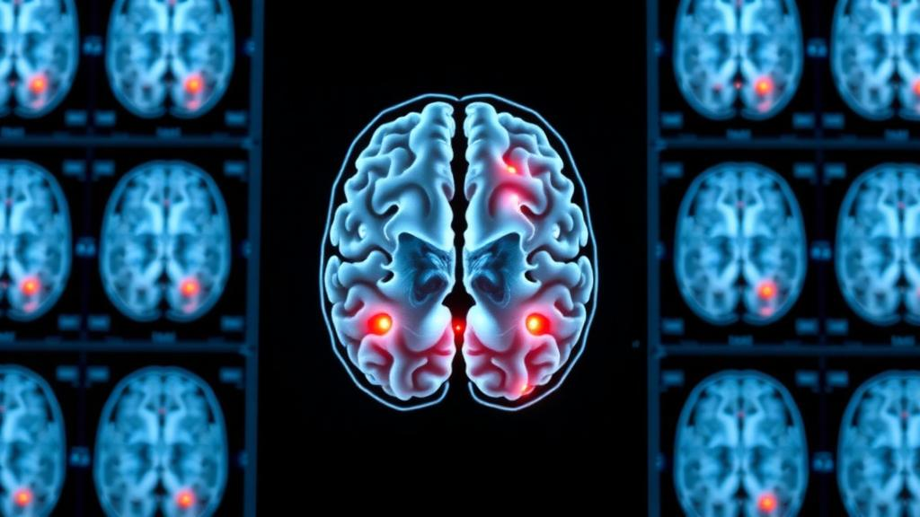
Perhaps the most clinically significant element is the framework’s insistence on showing its work. Deep networks are often dismissed as black boxes, and clinicians are rightly reluctant to trust a diagnosis they cannot interrogate. By applying Grad-CAM, which uses the gradients flowing into the final convolutional layer to produce heatmaps of influential image regions, the researchers demonstrated that their models consistently attended to tumor-relevant anatomy rather than spurious artifacts. Notably, the ensemble produced more focused attention maps than its individual members, while the simpler custom CNN spread its attention more broadly, mirroring its lower accuracy. The heatmaps offer radiologists visual evidence that the machine is looking at the same suspicious mass they are.
The authors are candid about the study’s limits. The classification and segmentation components were trained on two different public datasets, so the complete workflow has not yet been validated end-to-end on a single dataset carrying both labels and segmentation masks. All evaluation used public data, leaving generalisability to other institutions, scanners, and imaging protocols untested, and Grad-CAM, while informative, does not fully explain a network’s decision process. The framework has also not been deployed in real clinical workflows. Future work, the team says, will explore unified multi-task datasets, multi-centre validation, transformer-based and hybrid CNN-Transformer architectures, uncertainty estimation, and more advanced explainable AI techniques.
Even with those caveats, the study offers a compelling glimpse of where computer-aided diagnosis is heading: not a single oracle model, but an integrated pipeline that classifies, localizes, and justifies its conclusions. With the source code openly available on GitHub and both datasets publicly accessible, the work lowers the barrier for other groups to reproduce, critique, and extend it. As the authors put it in their conclusion, the results demonstrate the potential of deep learning systems to help radiologists achieve faster, more accurate, and more reliable brain tumor diagnosis — provided the machines can earn the same trust that human experts spend decades building.
Subject of Research: Deep learning framework for automated brain tumor classification, segmentation, and explainability in MRI
Article Title: Automated classification and segmentation of brain abnormalities using deep learning approaches in brain MRI
Article References: Nayak, R. R., Ramyashree, Raghavendra, S., & Anoop, B. N. (2026). Automated classification and segmentation of brain abnormalities using deep learning approaches in brain MRI. Discover Artificial Intelligence, 6(1), Article 1260. https://doi.org/10.1007/s44163-026-02253-5
Image Credits: AI Generated
DOI: 10.1007/s44163-026-02253-5
Keywords: brain tumor, MRI, deep learning, convolutional neural networks, ensemble learning, DenseNet121, InceptionV4, U-Net, image segmentation, Grad-CAM, explainable AI, computer-aided diagnosis
Cite Scienmag News
Cassandra Pierce. (October 16, 2026). AI Ensemble Reads Brain Scans With 98.5% Accuracy and Shows Its Work. Scienmag. https://scienmag.com/ai-ensemble-reads-brain-scans-with-98-5-accuracy-and-shows-its-work/
Cassandra Pierce. "AI Ensemble Reads Brain Scans With 98.5% Accuracy and Shows Its Work." Scienmag, 16 October 2026, https://scienmag.com/ai-ensemble-reads-brain-scans-with-98-5-accuracy-and-shows-its-work/. Accessed 16 October 2026.
Cassandra Pierce. "AI Ensemble Reads Brain Scans With 98.5% Accuracy and Shows Its Work." Scienmag. October 16, 2026. https://scienmag.com/ai-ensemble-reads-brain-scans-with-98-5-accuracy-and-shows-its-work/


