


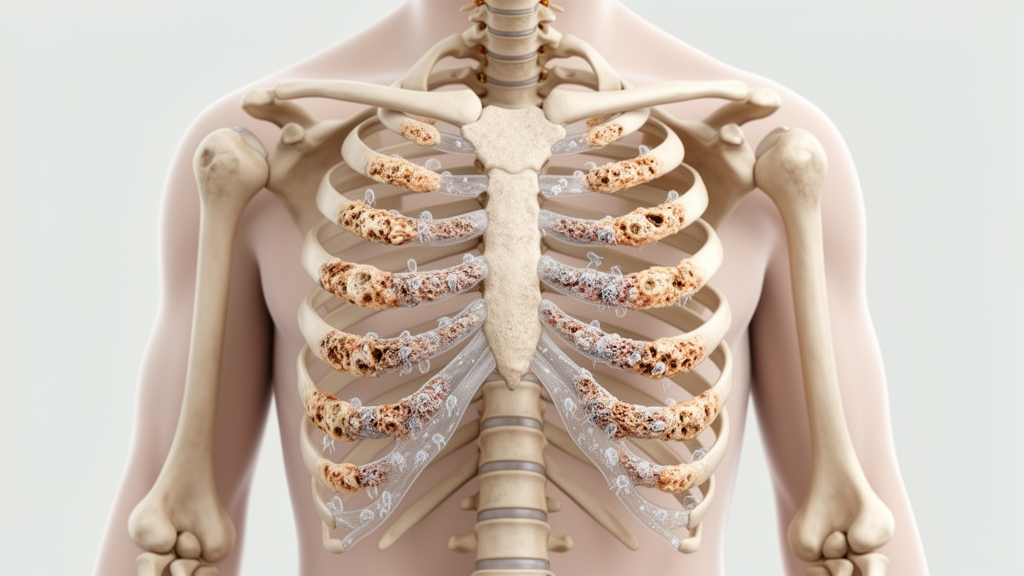
A 42-year-old woman with mild polyostotic fibrous dysplasia and precocious puberty had rib lesions newly detected on follow-up imaging despite the disorder’s activity typically declining in adulthood. The case also revealed an unusual bone-turnover profile: serum osteocalcin and alkaline phosphatase were within normal ranges, while procollagen type 1 N-terminal propeptide remained three to four times above the upper limit of normal. The authors report that permanent hypoparathyroidism may have influenced the pattern, but the single-patient study cannot establish that mechanism.
Fibrous dysplasia is a rare, mosaic skeletal disorder in which normal bone is replaced by fibro-osseous tissue. Disease activity typically peaks during childhood and diminishes by adulthood, and the extent and severity of skeletal involvement generally correlate with accelerated bone turnover. The patient had been diagnosed with mild polyostotic fibrous dysplasia 19 years before the new imaging findings.
Her history of precocious menarche at eight years of age later led clinicians to revise the diagnosis to fibrous dysplasia associated with McCune–Albright syndrome, abbreviated FD/MAS. Another defining feature of the patient’s medical history was permanent hypoparathyroidism. At age 31, she underwent total thyroidectomy for papillary thyroid cancer, and the operation was complicated by permanent hypoparathyroidism.
This condition became an important part of the case analysis because the measured bone-turnover markers did not follow the pattern generally associated with polyostotic fibrous dysplasia. During follow-up, 18F-sodium fluoride positron-emission tomography combined with computed tomography, or 18F-NaF PET/CT, identified rib lesions that had not been evident on an 18F-fluorodeoxyglucose PET/CT examination performed 19 years earlier when fibrous dysplasia was diagnosed. All of the newly detected skeletal foci showed morphological features consistent with fibrous dysplasia on the CT component of the NaF scan.
The report documents lesions recognized after a prolonged imaging interval; it does not establish when they arose or whether they represent renewed disease activity. Clinical and diagnostic checks did not offer an obvious alternative explanation for the rib findings. The patient had no clinical evidence of rib trauma and no biochemical or structural evidence of recurrent thyroid cancer.
The evidence therefore supports classifying the detected rib sites as additional skeletal manifestations consistent with fibrous dysplasia, while leaving open broader questions about their biological course in this adult case. Genetic testing supported the underlying diagnosis. Following target enrichment, next-generation sequencing of DNA extracted from the dysplastic right iliac bone identified the causal Arg201Cys variant in the GNAS gene.
The molecular result, combined with the earlier history of precocious puberty, established the revised classification as FD/MAS. Because the report concerns one person, however, the genetic finding characterizes this case rather than defining a general pattern for all adults with fibrous dysplasia. The most notable laboratory feature was the contrast among several bone-turnover indicators.
In polyostotic fibrous dysplasia, elevated markers would be expected in light of the association between skeletal involvement and accelerated bone turnover. In this patient, serum osteocalcin and alkaline phosphatase remained within their normal ranges. Procollagen type 1 N-terminal propeptide was the only marker reported as persistently elevated, staying three to four times above the upper limit of normal.
Authors suggested that this atypical profile could reflect the combined effects of FD/MAS and suppressed skeletal remodeling associated with hypoparathyroidism. That explanation remains a possibility rather than a demonstrated causal result. As a case report, the study offers a detailed clinical snapshot rather than a population-level estimate or controlled comparison.
Its scope is limited to one 42-year-old woman with mild polyostotic FD/MAS, permanent hypoparathyroidism, and additional rib foci detected on interval imaging. The authors’ principal conclusion is that skeletal lesions can become detectable in an adult case despite expected quiescence and that bone-turnover markers may display an unusual combination of normal and elevated results in the presence of FD/MAS and permanent hypoparathyroidism.
Subject of Research: Newly detected rib lesions and altered bone-turnover markers in adult fibrous dysplasia/McCune–Albright syndrome with hypoparathyroidism
Article Title: Adult fibrous dysplasia/McCune–Albright syndrome with hypoparathyroidism: additional skeletal lesions and an unusual bone turnover profile
Article References: Zajíčková, K., Včelák, J., Kuklíková, V., Černý, V., & Zogala, D. (2026). Adult fibrous dysplasia/McCune–Albright syndrome with hypoparathyroidism: additional skeletal lesions and an unusual bone turnover profile. BMC Endocrine Disorders. https://doi.org/10.1186/s12902-026-02687-z
Image Credits: AI Generated
DOI: 10.1186/s12902-026-02687-z
Keywords: fibrous dysplasia, McCune–Albright syndrome, hypoparathyroidism, rib lesions, bone turnover, PET/CT, GNAS variant, osteocalcin, procollagen type 1 N-terminal propeptide, Adult, fibrous, dysplasia
Cite Scienmag News
Ophelia Keating. (October 20, 2026). Adult fibrous dysplasia shows new rib lesions and unusual bone turnover. Scienmag. https://scienmag.com/adult-fibrous-dysplasia-shows-new-rib-lesions-and-unusual-bone-turnover/
Ophelia Keating. "Adult fibrous dysplasia shows new rib lesions and unusual bone turnover." Scienmag, 20 October 2026, https://scienmag.com/adult-fibrous-dysplasia-shows-new-rib-lesions-and-unusual-bone-turnover/. Accessed 20 October 2026.
Ophelia Keating. "Adult fibrous dysplasia shows new rib lesions and unusual bone turnover." Scienmag. October 20, 2026. https://scienmag.com/adult-fibrous-dysplasia-shows-new-rib-lesions-and-unusual-bone-turnover/


