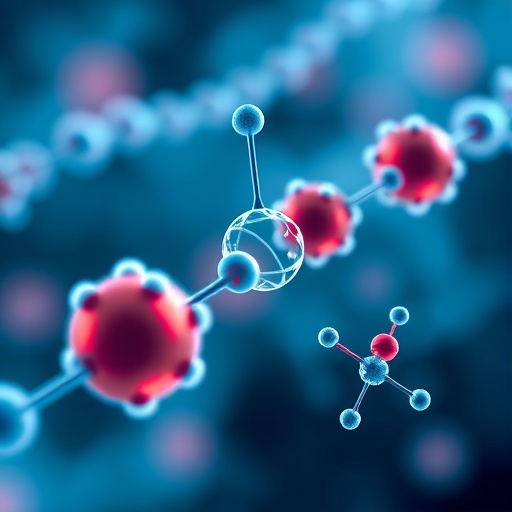
Lipid nanoparticles (LNPs) have emerged as pivotal carriers in the delivery of modern therapeutics, underpinning groundbreaking advances in cancer treatment, gene therapy, and vaccine technology. Long regarded as simple spherical vehicles ferrying molecular cargo across cellular landscapes, a recent collaborative study published in Nature Biotechnology shatters this simplistic paradigm. Researchers from the University of Pennsylvania, Brookhaven National Laboratory, and Waters Corporation have unveiled a complex landscape of LNP morphology, showing that these particles resemble more irregular “jelly beans” than perfect spheres. This revelation challenges fundamental assumptions about how LNPs function and offers new avenues to tailor these nanostructures for enhanced therapeutic precision.
Central to this revelation was the use of a trifecta of complementary biophysical techniques, enabling unprecedented scrutiny of LNP size, internal architecture, and cargo distribution—all while preserving the particles in their native, solution-phase environments. Sedimentation velocity analytical ultracentrifugation (SV-AUC), field-flow fractionation coupled with multi-angle light scattering (FFF-MALS), and size-exclusion chromatography integrated with synchrotron small-angle X-ray scattering (SEC-SAXS) were applied synergistically. Together, these methods deconvoluted the hydrodynamic profiles, density variations, and sub-nanometer structural organization within four benchmark LNP formulations, including those integral to COVID-19 vaccines and the FDA-approved Onpattro therapy.
This multifaceted approach marked a critical advance over prior studies that typically relied on isolated techniques—often freezing particles or tagging them with fluorescent markers—which inadvertently introduced artifacts or obscured structural heterogeneity. By circumventing these pitfalls, the team delineated variations not just between formulations but also among individual particles within the same batch. The findings reveal that LNPs are less uniform than previously thought, with shape and internal arrangement significantly influencing performance in biological systems.
Michael J. Mitchell, Associate Professor of Bioengineering at the University of Pennsylvania and a co-senior author of the study, likened the diversity to a fleet of specialized vehicles. “We no longer see LNPs as a homogenous model but rather a collection of distinct designs—akin to pickups, vans, and freight trucks tailored for varying therapeutic routes and targets,” Mitchell explained. This analogy encapsulates a shift towards recognizing the necessity for bespoke nanoparticle formulations optimized for specific tissues, cell types, and molecular payloads.
Kushol Gupta, Research Assistant Professor in Biochemistry and Biophysics and co-senior author, emphasized that understanding this complexity is not merely academic but foundational for clinical success. “Our work provides fundamental insights into how nanoparticle composition and architecture modulate biological interactions, potentially transforming the efficiency and specificity with which therapies reach their targets,” Gupta noted. The implications of this are profound: refined LNP design could accelerate the development of RNA therapies, enhance gene editing strategies, and reduce systemic side effects by ensuring precise delivery.
A particularly intriguing aspect of the research lies in the role of nanoparticle preparation methods. The study compared microfluidic mixing—a highly controlled, small-tube flow-driven process—and manual micropipetting. Though microfluidics generally yielded more uniform particles, micropipetting occasionally produced LNPs with superior functional profiles depending on the therapeutic context. This nuanced discovery highlights the importance of process engineering alongside chemical formulation, underscoring that small changes in manufacturing can dramatically impact nanoparticle efficacy and behavior in vivo.
The examination of how internal nanoparticle structure correlates with biological outcomes was further informed by testing across diverse models, including human T cells, cancerous cells, and animal studies. Doctoral researcher Hannah Yamagata found that no single LNP configuration was universally optimal. Instead, particle architecture needed to be matched judiciously to the target cell type or tissue environment for maximal delivery efficiency and therapeutic effect. This contextual dependency reiterates the fallacy of a one-size-fits-all approach, advocating for a paradigm of precision nanoparticle medicine tuned to the biological terrain.
Crucially, the study’s success was predicated on the synergy of academic, industrial, and national laboratory expertise. Waters Corporation provided sophisticated instrumentation for characterizing LNP size and drug load without disruption, while the National Synchrotron Light Source II at Brookhaven allowed nanoscale structural insights using intense X-ray beams. This cross-sector collaboration exemplifies the future of nanomedicine research, where deep specialization and shared resources converge to unravel complex biological phenomena.
Beyond technical sophistication, the work paves the path to predictive and rational LNP design, replacing the longstanding empirical “trial and error” methods that characterized the field. The integration of high-resolution structural data with biological performance metrics sets the stage for computational modeling and artificial intelligence approaches to anticipate how compositional tweaks and manufacturing conditions will influence therapeutic outcomes—potentially accelerating drug development timelines.
Importantly, while some of the analytical tools deployed (such as synchrotron radiation facilities) remain scarce, many laboratory techniques used for particle sizing and characterization are accessible to a broad range of researchers. The generation of shared, comprehensive data sets could catalyze an era of collaborative and data-driven nanoparticle engineering, democratizing advances for both academic inquiry and pharmaceutical innovation.
This study signifies a milestone, reframing lipid nanoparticles from passive carriers into complex functional devices whose architecture intimately governs their destiny within biological systems. As Mitchell concluded, “Our findings offer a roadmap for designing next-generation lipid nanoparticles with the precision and personalization akin to that of the drugs they carry, unlocking new therapeutic possibilities.”
Subject of Research: Cells
Article Title: Elucidating lipid nanoparticle properties and structure through biophysical analyses
News Publication Date: 23-Oct-2025
Web References: http://dx.doi.org/10.1038/s41587-025-02855-x
References: Nature Biotechnology, DOI: 10.1038/s41587-025-02855-x
Image Credits: Bella Ciervo
Keywords: Lipid nanoparticles, nanomedicine, RNA therapy, nanoparticle structure, biophysical characterization, synchrotron X-ray scattering, ultracentrifugation, microfluidics, therapeutic delivery, gene therapy, COVID-19 vaccine, nanoparticle heterogeneity
