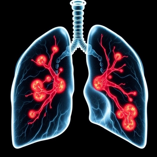
An innovative deep learning algorithm has emerged as a potential game-changer in the stratification of malignancy risks associated with pulmonary nodules. A recent study published in the esteemed journal, Radiology, indicates that this artificial intelligence-based tool not only excels in accurately identifying malignant growths but also significantly reduces the incidence of false positives. These findings are particularly crucial given the ongoing global battle against lung cancer, a disease responsible for more cancer-related fatalities than any other worldwide. The utilization of AI in medical diagnostics is forging new paths and, as illustrated in this study, has the power to refine how healthcare providers assess and manage lung nodules.
The landscape of lung cancer screening has long been fraught with challenges, predominantly concerning the ambiguous nature of pulmonary nodules—small, often oval-shaped growths which may or may not indicate malignancy. A prominent setback in past screening methodologies has been the high rates of false positives, which have imposed unnecessary anxiety upon patients and inflated healthcare costs due to excessive follow-up procedures. This situation has prompted the medical community to seek more reliable diagnostic models capable of discerning benign nodules from malignant ones with a greater degree of accuracy.
Traditionally, the assessment of lung nodule malignancy has leaned heavily on predefined parameters such as the size, type, and growth patterns of the nodules themselves. The Pan-Canadian Early Detection of Lung Cancer (PanCan) model represents a blend of patient and nodule characteristics to ascertain malignancy probabilities. Nevertheless, this probability-driven approach has its limitations, and the introduction of deep learning algorithms offers a compelling alternative that embraces fully data-driven predictions. The implications of such advancements could be profound, potentially altering current clinical practice guidelines and improving patient outcomes.
The retrospective study harnessed the power of a custom deep learning algorithm developed by researchers at Radboud University Medical Center, Nijmegen, Netherlands. Utilizing data from a robust national lung screening trial comprising over 16,000 nodules (including 1,249 malignant cases), this research endeavor aimed to create a model that could accurately estimate the malignancy risk associated with these growths. External validation employed CT scan data from several competing studies, further reinforcing the robustness of their outcomes.
Participants from the trial represented a diverse demographic, with a median age of 58 years and a majority (78%) being male. The extensive dataset allowed researchers to assess the algorithm’s efficacy across different cohorts, including both indeterminate nodules in the 5-15 mm size range and malignant nodules paired with size-matched benign equivalents. This targeted selection of indeterminate nodules is particularly pertinent, as these are the types that frequently require ongoing monitoring and can lead to substantial healthcare resource utilization if misclassified.
Compared against the PanCan model, the deep learning algorithm not only held its own but significantly outperformed it in multiple key metrics. In an analysis of the pooled cohort, AUC values—an essential indicator of a model’s diagnostic accuracy—revealed that the deep learning tool achieved scores of 0.98, 0.96, and 0.94 for cancers diagnosed at one year, two years, and throughout the screening process, respectively. The PanCan model, while respectable, lagged slightly behind with values of 0.98, 0.94, and 0.93, highlighting the emerging potential of AI methodologies in this critical area of medicine.
Particularly noteworthy is the performance of the algorithm when validating against indeterminate nodules—a group notorious for their diagnostic challenges. In this subset, the deep learning model achieved AUC scores of 0.95, 0.94, and 0.90 for short, medium, and long-term cancer predictions respectively, significantly outperforming the PanCan model’s scores of 0.91, 0.88, and 0.86. Such findings could usher in a new era where artificial intelligence can stratify risk with unprecedented accuracy, ultimately mitigating unnecessary procedures and enhancing patient management.
Of particular significance, the deep learning algorithm demonstrated a 39.4% relative reduction in false positives at 100% sensitivity for cancers diagnosed within one year. The model classified 68.1% of benign cases as low risk, in contrast to the PanCan model’s lower classification rate of just 47.4%. This stark delineation between the two models underlines the pressing need for integrating advanced AI tools into clinical practice, particularly to alleviate the burden often associated with false-positive results in lung cancer screening.
As the researchers and clinicians involved in the study advocate, the deep learning approach holds the promise of empowering radiologists in clinical decisions regarding follow-up imaging and management. However, researchers caution that while the preliminary results are compelling, further prospective validation is paramount to ascertain the clinical applicability of these tools. Future investigations must guide the method’s implementation in real-world settings, ultimately refining the lung cancer screening paradigm.
In this multifaceted exploration of pulmonary nodule malignancy risk stratification, the collaborative effort led by Dr. Noa Antonissen and an extensive cohort of researchers signals a pivotal juncture in the intersection of artificial intelligence and healthcare. Backed by entities such as the Dutch Cancer Society and Siemens Healthineers, this research is a testament to the promise that lies at the convergence of technology and medicine.
The future of lung cancer screening may soon witness a transformation characterized by enhanced accuracy and reduced anxiety for patients. With the commitment to advancing deep learning methodologies, researchers may be on the precipice of routinely utilizing these sophisticated algorithms as standard practice tools. Through continued innovation and validation, the hope is to not only enhance screening efficacy but to also ultimately save lives by enabling earlier detection of lung malignancies.
With a growing emphasis on integrating artificial intelligence into clinical workflows, the findings of this study will likely initiate broader discussions on how healthcare institutions can adapt to leverage emerging technologies effectively. As healthcare continues to navigate overarching challenges, persistent efforts in refining diagnostic tools could lead to a future where lung nodules no longer represent a source of fear, but rather an opportunity for proactive health management.
The study showcasing the deep learning model’s effectiveness stands at the forefront of a new era in medical diagnostics. By addressing the intricate challenges presented by lung cancer screening, researchers are paving the way for improved patient outcomes while setting a precedent for future innovations in oncology. From bolstering screening accuracy to enhancing patient confidence, the contributions of leaders in AI research like Dr. Antonissen signal a monumental stride toward realizing a healthier future for those at risk of lung cancer.
In summary, the continuing evolution of lung nodule malignancy risk assessment through deep learning represents an inspiring chapter in the medical landscape. The marriage of technology with healthcare holds tremendous potential, enabling groundbreaking solutions that promise to enhance diagnostic accuracy and, consequently, patient care in lung cancer screening.
Subject of Research: Lung Cancer Screening and Deep Learning Algorithms
Article Title: AI Deep Learning Tool Revolutionizes Lung Nodule Malignancy Risk Assessment
News Publication Date: September 16, 2025
Web References: Radiology Journal
References: N/A
Image Credits: Radiological Society of North America (RSNA)
Keywords
Lung cancer, Artificial intelligence, Cancer screening, Mortality rates


