In a groundbreaking advancement poised to reshape oncological prognostics, a recent study has unveiled a pioneering clinical model designed to accurately identify gastroenteropancreatic neuroendocrine tumor (GEP-NET) patients who harbor a high burden of metastatic liver tumors. Traditionally, the evaluation of liver tumor burden (LTB) has relied heavily on intricate radiologic and functional imaging techniques, which, despite their precision, present significant logistical challenges in routine clinical settings. This innovation, spearheaded by a team of researchers, offers a streamlined, clinicopathology-based approach that leverages commonly available biochemical markers, marking a significant leap toward personalized cancer management.
Gastroenteropancreatic neuroendocrine tumors comprise a heterogeneous group of neoplasms originating from the hormone-producing cells of the gastroenteric and pancreatic systems. These tumors, while often indolent, have the propensity to metastasize, with the liver representing the most frequent and clinically consequential site of secondary involvement. The metastatic liver tumor burden is a critical determinant of patient prognosis, influencing survival outcomes and guiding therapeutic decision-making. Despite its importance, standardized methods for assessing LTB have remained cumbersome, necessitating this novel approach.
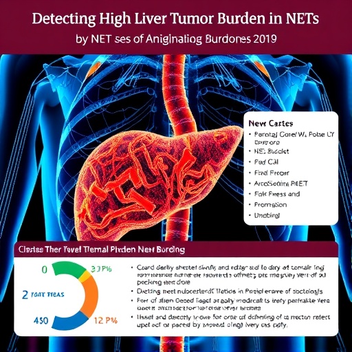
Central to this study is the quantification of liver tumor burden using the advanced imaging modality of ^68Ga-DOTANOC PET/CT scanning. This radiotracer-based technique offers exquisite sensitivity in detecting neuroendocrine tumor lesions but demands resources and expertise that are not ubiquitously accessible. Hence, the researchers embarked on an ambitious endeavor to circumvent these limitations by identifying surrogate clinicopathological markers that can predict LTB with comparable accuracy.
The research cohort encompassed 200 patients diagnosed with well-differentiated GEP-NETs. These subjects underwent thorough clinical evaluation, with serum levels of liver enzymes such as gamma-glutamyltransferase (GGT) and lactate dehydrogenase (LDH), along with tumor biomarkers inclusive of neuron-specific enolase (NSE), measured within a narrow temporal window prior to PET/CT imaging. A critical histopathological parameter, the Ki-67 proliferation index, was also integrated, reflecting the tumor’s intrinsic growth dynamic.
Employing an advanced statistical technique known as Least Absolute Shrinkage and Selection Operator (LASSO) regression, the investigators meticulously sifted through numerous potential predictors to isolate those variables most significantly associated with high LTB. The final predictive quartet emerged as Ki-67 index, GGT, LDH, and NSE, each contributing distinct pathophysiological insights. Ki-67 gauges cellular proliferation, GGT and LDH reflect hepatocellular and systemic metabolic distress, while NSE serves as a surrogate marker for neuroendocrine activity.
This methodological rigor culminated in the construction of a nomogram—a graphical computational tool—that translates these variables into a quantifiable risk score, enabling clinicians to estimate the likelihood of a patient exhibiting a high liver tumor burden. The nomogram’s performance was robust, achieving an area under the curve (AUC) of approximately 0.78 in both training and validation cohorts, indicative of high discriminative capability and model generalizability.
The practical implications of this predictive tool are profound. By stratifying patients according to their nomogram-derived total points into high and low LTB groups, physicians can now anticipate clinical outcomes with greater precision. Notably, those classified within the high LTB category (total points ≥ 26.2) demonstrated significantly worse overall survival, underscoring the nomogram’s prognostic validity and potential to inform aggressive versus conservative management strategies.
From a therapeutic standpoint, early and accurate identification of patients with extensive liver involvement could prioritize them for more intensive interventions such as peptide receptor radionuclide therapy (PRRT), hepatic arterial embolization, or systemic chemotherapy. Conversely, patients with low tumor burden may be spared from aggressive treatments, mitigating toxicity and preserving quality of life. This nuanced risk-adapted approach epitomizes the ideals of personalized medicine.
Beyond prognostication, the utilization of routinely accessible blood tests as the foundation of this model enhances its applicability across diverse healthcare settings, including those with limited access to advanced imaging. Such democratization of diagnostic capability is pivotal in bridging disparities in cancer care, particularly in resource-constrained environments.
The study also adds valuable insight into the biological underpinnings of GEP-NET metastasis. The correlation of elevated liver enzymes with tumor burden may reflect the hepatic parenchymal response to metastatic infiltration, while elevated NSE underscores the neuroendocrine phenotype’s role in disease progression. These findings not only bolster the biological plausibility of the model but also invite further exploration into the mechanistic pathways of liver metastasis.
Moreover, the inclusion of the Ki-67 index, a well-established proliferative marker, reiterates its critical place in neuroendocrine tumor grading and outcome prediction. Integrating this parameter with biochemical markers bridges histopathological assessment and systemic disease markers, fostering a comprehensive view of tumor behavior.
The retrospective nature of the study, encompassing a substantial patient cohort, lends weight to the findings, though future prospective validation studies will be essential to cement the model’s role in clinical practice. Additionally, expanding this predictive framework to include genomic or molecular profiling could further refine its accuracy and uncover novel therapeutic targets.
In an era where artificial intelligence and machine learning increasingly intersect with medicine, the application of LASSO regression exemplifies how sophisticated analytical methods can distill complex biological data into clinically actionable formats. This synergy between technology and human expertise promises to accelerate the pace of oncological innovation.
This clinical model’s offering is timely, addressing the escalating incidence of GEP-NETs and the pressing need for effective, accessible tools to guide clinical decision-making. As therapeutic options expand and evolve, accurate tumor burden estimation will remain a cornerstone of optimal patient management.
Ultimately, this research heralds a paradigm shift, transforming the evaluation of metastatic neuroendocrine tumors from an exclusive reliance on imaging into a multi-modal, biomarker-driven assessment. By enabling early, precise identification of high-risk patients, it paves the way for tailored treatment approaches that can improve survival and quality of life.
Clinicians, researchers, and patients alike stand to benefit from this innovation, illustrative of how rigorous scientific inquiry can translate into tangible health advances. As this nomogram gains traction, it may serve as a template for analogous predictive tools across cancer subtypes, fostering a new standard of precision oncology.
The integration of clinicopathological data into a predictive tool underscores the evolving landscape of cancer diagnostics, where bedside-to-bench and bench-to-bedside knowledge cycles are increasingly intertwined. This holistic approach highlights the power of multidisciplinary collaboration in addressing complex clinical challenges.
In summary, the development of this nomogram by Jumai and colleagues embodies a significant stride forward in managing gastroenteropancreatic neuroendocrine tumors. By harnessing readily accessible clinicopathological markers, the model promises to streamline risk stratification, personalize treatment, and ultimately improve patient outcomes in this challenging disease realm.
Subject of Research: Prediction of liver tumor burden in gastroenteropancreatic neuroendocrine tumor patients using clinicopathological markers.
Article Title: Identification of gastroenteropancreatic neuroendocrine tumor patients with high liver tumor burden based on clinicopathological features.
Article References:
Jumai, N., Chen, L., Lin, X. et al. Identification of gastroenteropancreatic neuroendocrine tumor patients with high liver tumor burden based on clinicopathological features. BMC Cancer 25, 1217 (2025). https://doi.org/10.1186/s12885-025-14535-9
Image Credits: Scienmag.com
DOI: https://doi.org/10.1186/s12885-025-14535-9
