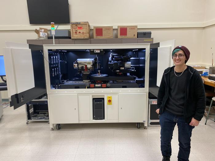
Interdisciplinary researchers in Illinois, the U.S. and around the world can advance their projects with the Beckman Institute for Advanced Science and Technology’s new Zeiss Xradia 630 Versa micro-CT scanner, the first of its kind with life science applications in the U.S.

Credit: Jenna Kurtzweil, Beckman Institute Communications Office.
Interdisciplinary researchers in Illinois, the U.S. and around the world can advance their projects with the Beckman Institute for Advanced Science and Technology’s new Zeiss Xradia 630 Versa micro-CT scanner, the first of its kind with life science applications in the U.S.
Computed tomography, or CT, is an imaging technique that involves capturing a series of cross-sectional X-ray scans of an object or sample — be it a material like concrete or a biological sample like an insect or human body. Stacked on top of one another, the images non-invasively reconstruct the subject in 3D from the inside out. Microscopic computed tomography, or micro-CT, helps researchers reconstruct subjects as tiny and delicate as collagen or insect antennae.
Most micro-CT scanners use only X-rays, which are invisible to the human eye, but Beckman’s new model also incorporates visible through a process called scintillation. The 630 Versa can also scan small subsections of large materials while maintaining a maximum imaging resolution of 40 nanometers, 2,500 times narrower than the width of a human hair.
“With building materials, hairline cracks are the start of where things go catastrophically wrong, and the micro-CT scanner at the Beckman Institute can see those cracks,” said Beckman microscopist T. Josek. “We’re providing the opportunity for researchers at the University of Illinois and beyond to start here and start small — see how a material behaves and what it looks like, and then scale up.”
Researchers can use the compression/tensile stage to stretch, bend and compress their samples while scrutinizing them up close — a technique called in situ mechanical testing. The Versa 630 can also do diffraction contrast tomography, a technique wherein researchers can determine a material’s grain orientation by tracking how X-rays bend as they pass through it.
The scanner was installed in January 2024 and is the first 630 model for life science applications in the U.S. In addition to the Beckman Institute, the Illinois Materials Research Laboratory, The Grainger College of Engineering and the Office for the Vice Chancellor of Research and Innovation committed funds to support its purchase.
The Beckman Microscopy Suite team is focused on finding projects on campus and beyond that might benefit from the Versa 630 and are providing comprehensive trainings for those new to the technology. Carl Zeiss Microscopy, the scanner’s manufacturer, is integral to these efforts.
“We at ZEISS are thrilled to be partnering with the Beckman Institute Microscopy Suite to deliver this flexible and intuitive system for X-ray microscopy,” said Aubrey Funke, Carl Zeiss Microscopy Product Marketing Manager for Life Science EM/XRM. “The ability to image your sample, be it organic or material, becomes a less intimidating process on the ZEISS Xradia Versa 630. With high powered-resolution and expanded ease-of-use for all skill levels, the Versa 630 is ideal for diverse research capabilities across a spectrum of applications, including life science, electronics, materials research, and many more. We look forward to working with the Beckman Institute Microscopy Suite to support many diverse research projects.”
Researchers at the University of Illinois and beyond are encouraged to use the 630 Versa micro-CT scanner. For more information or to use the micro-CT scanner, please visit the Beckman Institute Microscopy Suite website at https://itg.beckman.illinois.edu/microscopy_suite.


