


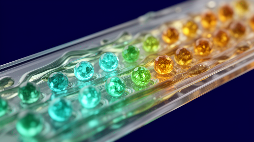
Quantum dot biosensors have emerged as a promising platform for biomedical diagnostics, particularly for early disease detection where conventional immunoassays often struggle with low sensitivity, photobleaching, and poor optical penetration in biological tissues. A recent review published in Discover Sensors highlights recent advances in these nanoscale semiconductor sensors, emphasizing their operating principles, analytical performance, and utility in detecting pathogens, cancer biomarkers, metabolites, and other clinically relevant targets. The study provides a mechanistic and integrated perspective by linking quantum dot surface engineering with sensing mechanisms and their clinical translational relevance, offering a unified framework for next-generation diagnostic development.
Quantum dots are nanoscale semiconductor nanocrystals, typically 2 to 10 nanometers in diameter, consisting of an inorganic core stabilized by organic ligands or polymeric surface molecules. Their fundamental optical behavior arises from quantum confinement effects, where the electronic band gap changes based on the nanocrystal size. When the diameter becomes smaller than the exciton Bohr radius, the band gap increases, resulting in blue-shifted emission; conversely, larger quantum dots exhibit narrower bandgaps and red-shifted emission. This size-dependent tunability allows researchers to control emission from the ultraviolet to the near-infrared range by simply modulating particle size, a feature that is highly attractive for biosensing applications due to their broad absorption spectra and narrow emission profiles.
The exceptional photostability and high quantum yield of quantum dots enable reliable detection even at ultralow biomarker levels, overcoming the limitations of traditional organic dyes that often suffer from photobleaching. Their surface chemistry can be precisely engineered to attach antibodies, aptamers, peptides, or nucleic acids, supporting a wide range of selective and multiplexed sensing formats. Near-infrared emitting quantum dots further enhance clinical applicability by reducing tissue autofluorescence and improving in vivo imaging depth, which is critical for real-time molecular tracking and deep-tissue diagnostics. This capability allows for simultaneous imaging of multiple biomarkers, facilitating comprehensive disease profiling with high precision.
Fluorescence-based biosensing relies on the superior optical properties of quantum dots, which exhibit higher fluorescence intensity and longer emission lifetimes compared to conventional probes. The fundamental detection mechanism is typically based on fluorescence quenching, where the emitted signal decreases due to specific molecular interactions between quantum dots and a quencher. Quenching can occur through static pathways, dynamic collisional processes, inner filter effects, or Förster resonance energy transfer. For instance, a ratiometric fluorescence sensor using cadmium telluride sulfide quantum dots achieved a detection limit of 0.78 micromolar for ascorbic acid, demonstrating reliable performance in vitamin C tablet analysis with a linear range of 1 to 500 micromolar.
Photoelectrochemical biosensors offer another avenue for sensitive detection by relying on the light-induced generation and separation of electron-hole pairs in semiconductor photoelectrodes. Upon illumination, absorbed photons create charge carriers that participate in interfacial redox reactions, producing a measurable photocurrent. Biorecognition events, such as antigen-antibody binding, alter surface states and charge-transfer efficiency, leading to a detectable change in this photocurrent. A recent study developed an ultrasensitive photoelectrochemical immunosensor for cardiac troponin I detection using a dual inhibition strategy, achieving an exceptionally low detection limit of 6.7 femtograms per milliliter. This approach demonstrated admirable stability and reproducibility, providing a novel strategy for high-performance immunoassay development.
Electrochemiluminescence biosensors generate light through electrochemical reactions without requiring optical excitation, which can reduce background noise in complex biological matrices. In these systems, electrochemical processes produce excited-state quantum dots that emit light, often utilizing a coreactant pathway where reactive intermediates transfer electrons to the quantum dots. An electrochemiluminescence biosensor using cobalt-doped cadmium telluride quantum dots detected human carcinoembryonic antigen with a linear range of 1 femtogram to 10 picograms per milliliter and a detection limit of 0.2 femtograms per milliliter. Another platform using sulfur quantum dots achieved a detection limit of 6.67 attomolar for microRNA-21, highlighting the extreme sensitivity possible with these non-optical excitation methods.
Surface modification is essential for improving the stability, solubility, and biocompatibility of quantum dots in physiological environments. Various strategies, including covalent conjugation, ligand exchange, and electrostatic interaction, allow for the precise attachment of biomolecules to the quantum dot surface. Covalent bonding through EDC/NHS chemistry provides a stable method for attaching biomolecules, while ligand exchange enables the transfer of hydrophobic quantum dots into aqueous media. These modifications enhance the performance and applicability of quantum dots in biological systems by preventing aggregation and nonspecific interactions, thereby enabling selective binding and precise imaging of specific cells or biomolecules.
Despite their advantages, the clinical translation of quantum dot biosensors is hindered by concerns regarding toxicity and biological safety. The primary source of toxicity in conventional quantum dots arises from their heavy-metal cores, such as cadmium selenide and cadmium telluride, which can degrade and release metal ions under intracellular oxidative conditions. Such ion leakage can disrupt cellular functions and induce apoptosis. To mitigate these risks, researchers have developed cadmium-free quantum dots, such as indium phosphide and silicon systems, which show markedly reduced toxicity and comparable optical properties. Additionally, surface passivation with zinc sulfide shells or biocompatible coatings like polyethylene glycol can reduce cytotoxicity and improve long-term stability in biological environments.
The integration of artificial intelligence and machine learning with quantum dot-based sensing platforms has emerged as a transformative approach to enhance sensor performance and data interpretation. Machine learning techniques leverage experimental datasets to extract patterns and predict complex responses, enabling high-throughput and accurate biosensing applications. For example, a deep learning-based platform for interleukin-6 detection achieved a limit of detection as low as 3.1 femtomolar, while another system used optimized principal component analysis to simultaneously detect four gastrointestinal tumor markers with high classification accuracy. These AI-driven approaches improve diagnostic accuracy and facilitate real-time monitoring, bridging the gap between laboratory research and practical clinical applications.
Looking forward, the future of quantum dot biodiagnostics lies in the development of robust, biocompatible, and scalable sensing platforms. Advancements in heavy-metal-free quantum dots and improved ligand engineering are expected to minimize toxicity and enhance long-term stability. The synthesis of highly luminescent near-infrared quantum dots will further enable deep-tissue imaging with minimal autofluorescence, allowing enhanced diagnostic precision in applications such as tumor imaging and traumatic brain injury detection. As these technologies are integrated into miniaturized diagnostic systems and portable point-of-care platforms, quantum dot biosensors are poised to play a significant role in precision medicine and personalized biomedical interventions.
Beyond metabolic assays, quantum dot biosensors have demonstrated notable utility across viral and bacterial diagnostic formats. A graphene oxide-coated nanopaper immunosensor employing antibody-functionalized quantum dots demonstrated rapid pathogen detection through a quenching-recovery mechanism, identifying Escherichia coli at 55 to 70 colony-forming units per milliliter or gram and human immunoglobulin G at 1.60 nanograms per milliliter across serum, poultry, and water without wash steps. In viral sensing, cadmium telluride quantum dots conjugated to avian influenza virus H5N1 antibodies yielded a limit of detection of approximately 1.5 × 10⁻⁴ micrograms per milliliter in sandwich assays. For human immunodeficiency virus detection, a strip integrating strand displacement amplification with two hairpin DNA sequences attained a detection limit of 0.76 picomolar. Additionally, a room-temperature-stable quantum dot sensor targeted SARS-CoV-2 genetic material in clinical samples within one hour, achieving a detection limit of one virus per milliliter at under $1.50 per test.
Quantum dot platforms also show efficacy for oncological screening and acute tissue injury monitoring. Because cancerous cells contain up to four-fold higher intracellular glutathione concentrations than normal cells, an inner filter effect biosensor coupling silver sulfide quantum dots with cobalt oxyhydroxide nanoflakes monitored glutathione via redox interactions releasing cobalt ions. For circulating tumor cell identification, copper-based near-infrared quantum dots linked to EpCAM antibodies achieved a detection limit of 12 MCF-7 breast cancer cells per well. In addition, an aptamer platform using silver sulfide nanodots and hybrid cell membrane-coated magnetic nanoparticles demonstrated 97.63 percent capture efficiency, detecting 6 to 10 circulating tumor cells per milliliter in patient samples with zero detection in healthy controls. For acute trauma, a quenched silver sulfide nanoprobe operating in the second near-infrared window targeted inflamed vascular endothelium via VCAM1-mediated endocytosis, restoring fluorescence upon reaction with peroxynitrite to track traumatic brain injury progression.
Alternative transduction platforms further expand diagnostic capacity beyond standard fluorescence. In photoelectrochemical sensing, combining p-type copper indium disulfide quantum dots with upconversion nanoparticles produced both a cathodic photocurrent and a visible green emission signal for prostate-specific antigen detection, achieving a limit of detection of 0.03 picograms per milliliter. In electrochemiluminescence, a potential-resolved immunosensor integrating luminol, carbon quantum dots, and cadmium sulfide quantum dots on gold nanoparticles simultaneously quantified three latent tuberculosis infection biomarkers, interleukin-2, tumor necrosis factor-alpha, and interferon-gamma, across a linear range of 1.6 to 200 picograms per milliliter. Furthermore, spin-based quantum dot sensors compatible with spintronics and CMOS architecture demonstrated that circularly polarized light produced a two-fold enhancement in spin polarization and a ten-fold increase in DNA hybridization sensitivity relative to unilluminated conditions.
To address stability and excretion, carbon quantum dots offer distinct resilience and clearance profiles. Carbon quantum dots maintain emission across pH 5 to 11 and resist interference from common physiological cations including sodium, potassium, calcium, and magnesium. In biodistribution evaluations, carbon dots undergo hepatobiliary clearance and fecal excretion. Similarly, engineering nanocrystals below a hydrodynamic diameter of 5 to 6 nanometers promotes renal clearance, helping avoid long-term organ accumulation.
Subject of Research: Quantum dot biosensors for sensitive and multiplex biomedical diagnostics
Article Title: Quantum dot biosensors for highly sensitive and multiplex detection in biomedical diagnostics
Article References: Patra, S., & Shand, H. (2026). Quantum dot biosensors for highly sensitive and multiplex detection in biomedical diagnostics. Discover Sensors, 2(1), Article 32. https://doi.org/10.1007/s44397-026-00064-y
Image Credits: AI Generated
DOI: 10.1007/s44397-026-00064-y
Keywords: quantum dots, biosensors, biomedical diagnostics, fluorescence, photoelectrochemical, electrochemiluminescence, nanotechnology, biocompatibility, machine learning, near-infrared imaging, Quantum, highly
Cite Scienmag News
Katie Riggs. (November 11, 2026). Quantum Dot Biosensors Advance Sensitive Multiplex Diagnostics. Scienmag. https://scienmag.com/quantum-dot-biosensors-advance-sensitive-multiplex-diagnostics/
Katie Riggs. "Quantum Dot Biosensors Advance Sensitive Multiplex Diagnostics." Scienmag, 11 November 2026, https://scienmag.com/quantum-dot-biosensors-advance-sensitive-multiplex-diagnostics/. Accessed 11 November 2026.
Katie Riggs. "Quantum Dot Biosensors Advance Sensitive Multiplex Diagnostics." Scienmag. November 11, 2026. https://scienmag.com/quantum-dot-biosensors-advance-sensitive-multiplex-diagnostics/




