
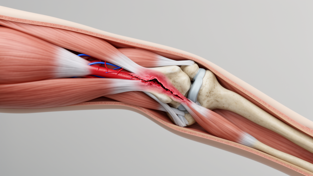
A case report published in Discover Medicine in September 2026 describes a rare clinical scenario in which a complete disconnection of the brachial artery was initially misdiagnosed as a simple elbow dislocation. The study details the experience of a 36-year-old male patient who sustained an accidental fall, resulting in pain, restricted movement, and significant discomfort in the left elbow. Although the limb appeared well-perfused upon admission, the patient subsequently developed a delayed vascular crisis, highlighting the deceptive nature of this specific injury pattern. The authors emphasize that such occult vascular injuries are highly susceptible to missed diagnoses due to the absence of typical ischemic signs in the early stages of presentation.
The diagnostic challenge in this case stemmed from the complex anatomy of the elbow and the presence of a robust collateral vascular network. According to the report, the initial physical examination indicated swelling and deformity of the left elbow joint, limited range of motion, and compromised peripheral blood supply. However, the nail bed was moderately full, and muscle strength and tone in the limbs were found to be normal. This deceptive clinical picture, characterized by a pink appearance of the limb, created a false sense of security for the clinicians. The preliminary diagnosis included dislocation and fracture of the left elbow joint, along with possible vascular and nerve injury, but the complete arterial disconnection was not immediately apparent.
Imaging studies played a crucial role in the initial assessment and surgical planning. X-ray and computed tomography (CT) of the elbow joint revealed the dislocation of the left elbow joint accompanied by avulsion fractures of the lateral condyle of the humerus and the olecranon of the ulna. Pre-reduction CT 3D reconstruction was particularly important for delineating the relationship between the bony fragments and the joint displacement. Despite the severity of the bony injury, the initial physical examination continued to show a well-perfused limb, underscoring the risk of missing vascular injury based on appearance alone. The imaging findings confirmed the posterior dislocation and the associated fractures, but did not initially reveal the extent of the vascular damage.
In the emergency department, manual reduction of the elbow dislocation was performed under brachial plexus nerve block anesthesia. Subsequent X-ray examination confirmed the successful reduction of the joint structures. Following immobilization of the left upper limb with a splint, the patient was transferred back to the ward. However, a few hours later, the patient complained of decreased sensation in the left hand. Physical evaluation revealed elbow swelling, absence of palpable radial artery pulse in the left wrist, and pink discoloration of the left fingertip. These signs indicated a change in the vascular status, prompting further investigation.
Immediate vascular ultrasound identified blood accumulation in the elbow joint, raising concerns of vascular injury and partial embolism. The ultrasound findings illustrated the deceptive nature of the brachial artery injury, showing retrograde collateral flow that maintained distal perfusion despite the proximal injury. Grayscale imaging revealed intraluminal hypoechoic thrombus, indicating occult occlusion, while color Doppler demonstrated incomplete vessel filling suggestive of stenosis or dissection. A transverse view showed perivascular hematoma compressing the vessel lumen, and evidence of severe soft tissue disruption and edema contributing to external vascular compression. These images collectively demonstrated how robust collateral circulation and partial patency could mask a critical arterial injury.
Emergency exploration was performed based on the ultrasound findings. Intraoperative findings confirmed the preoperative suspicion of vascular disconnection, revealing a complete transection of both the brachial artery and vein with lacerated ends and intravascular thrombosis. The median nerve sustained only epineurial contusion, and there was a partial tear of the left biceps brachii and elbow joint capsule ligament. No fasciotomy was performed as the compartment pressure remained within normal limits. Following thrombectomy, the mobilized brachial vein was of sufficient length to allow for primary end-to-end anastomosis. The elbow joint capsule ligament was first repaired using absorbable sutures, followed by the venous repair using an 8–0 Prolene suture.
However, after debridement, a 4-cm defect in the brachial artery necessitated reconstruction using an interposition vein graft harvested from the medial aspect of the left knee. The biceps brachii was then sutured, and the elbow was immobilized using a modular external fixator at 20° of flexion to prevent compression of the brachial artery anastomosis. The total ischemia time, from admission to intraoperative revascularization, was approximately 18 hours. Postoperatively, a strict anticoagulation protocol involving low molecular weight heparin combined with cilostazol was administered to prevent further thrombotic events. This initial surgical intervention aimed to restore blood flow and stabilize the joint.
One day post-operation, the patient complained of significant pain in the left elbow, yet noted an improvement in sensation in the left hand. Physical examination indicated a rosy peripheral color in the left hand, sensitive capillary response, and weaker radial artery pulsation compared to pre-operation. Color Doppler ultrasound assessment of the left upper limb vessels displayed unclear intracavascular fluid area in the distal segment of the left brachial artery at the site of the transplanted vessel, with no apparent blood flow signal. Flow velocity reduction was observed in the middle and lower segments of the left brachial artery, ulnar artery, and radial artery. These findings suggested a complication with the initial repair.
Emergency vascular crisis exploration and repair surgery was conducted in the emergency department under general anesthesia. Upon suture removal, surgical exploration identified the exact cause of the complication, which was found to be extensive thrombosis involving the proximal brachial artery, the proximal anastomosis, the interposition vein graft segment, and the distal anastomosis. Under microscopic magnification, a thrombectomy was performed, and the thrombosed vascular segments were completely resected. After releasing the vascular clamps, satisfactory pulsatile flow was observed from both the proximal and distal ends. A great saphenous vein was then harvested from the left thigh to reconstruct the brachial artery defect.
After adjusting the tension, the vessel ends were anastomosed end-to-end using 8–0 Prolene, and the adventitia was appropriately loosened to prevent stenosis. Post-revascularization, the radial artery pulse returned to normal, the fingertips appeared pink with brisk capillary refill, and the hand exhibited normal temperature and tension. The patient’s condition stabilized postoperatively, and strict bed rest was prescribed to mitigate swelling and alleviate pain. Following the subsequent physical therapy regimen, the patient experienced a gradual return of sensation in the left hand by the second postoperative day, alongside significant relief of elbow pain.
Two weeks postoperatively, sutures were removed, and supervised elbow rehabilitation exercises were initiated under the guidance of a physical therapist. At the 1-month follow-up, the external fixator was removed, and the patient demonstrated improved elbow joint mobility. Currently, the patient has achieved substantial functional recovery. The Lomb function was favourable in 2 years follow-up, with range of motion exercises demonstrating normal flexion, extension, pronation, and supination. The video accompanying the follow-up shows dynamic recovery, indicating a successful long-term outcome for this complex injury.
The discussion highlights the dynamic nature of the “painless” limb, noting that the initial pink appearance was maintained by a robust collateral network, specifically the Superior Ulnar Collateral Artery and Inferior Ulnar Collateral Artery. This compensatory mechanism failed post-reduction, likely due to the loss of the tamponade effect on the vascular tear by fracture fragments or the expansion of a hematoma, triggering delayed vascular compromise. The primary reason for the diagnostic delay lies in the complex anatomy of the elbow, where the rich collateral vascular network allows for adequate tissue perfusion even in the event of a complete transection of the main brachial artery. This results in a deceptive clinical triad of palpable pulses due to collateral flow, normal capillary refill in the early phase, and absence of gross pallor.
Furthermore, the patient’s history of smoking posed a synergistic risk, with nicotine-induced endothelial dysfunction likely accelerating thrombosis post-operatively and contributing to the graft failure and the subsequent crisis. The case validates the necessity of performing computed tomography angiography (CTA) in high-risk patients, as the traditional “6P” signs are reliable for overt ischemia but fail in occult injuries. For patients presenting with elbow fracture-dislocation with avulsion fragments, neurological symptoms out of proportion to the injury, or a history of smoking or vascular risk factors, the authors recommend bypassing duplex ultrasound and proceeding directly to CTA. Unlike ultrasound, which is operator-dependent and can be misled by collaterals, CTA provides a definitive anatomical roadmap and remains the gold standard for diagnosing vascular injuries.
Based on this experience, the authors propose a phased management strategy. Preoperatively, high-risk patients require immediate CTA. Intraoperatively, if a hematoma or neurological deficit is present, full exposure of the brachial artery is required, with thorough debridement. Postoperatively, implementation of strict quantitative monitoring, such as pulse grading and skin temperature differentials, during the first 24 hours is essential to detect delayed thrombosis. Protocolized management is key to limb salvage in these complex injuries. In conclusion, concealed brachial artery disconnection is a life-threatening diagnostic pitfall, and clinicians must maintain a high index of suspicion in elbow fracture-dislocations with neurological symptoms or risk factors. Prompt CTA and dynamic postoperative monitoring are the cornerstones of preventing limb loss in these cases.
Subject of Research: Diagnostic pitfall of concealed brachial artery disconnection in elbow trauma
Article Title: Navigating the diagnostic pitfall of concealed brachial artery disconnection masquerading as simple elbow dislocation
Article References: Gao, L., Guo, S., Chao, Y., & Liu, X. (2026). Navigating the diagnostic pitfall of concealed brachial artery disconnection masquerading as simple elbow dislocation. Discover Medicine, 3(1), Article 127. https://doi.org/10.1007/s44337-026-00689-3
Image Credits: AI Generated
DOI: 10.1007/s44337-026-00689-3
Keywords: elbow dislocation, brachial artery, vascular injury, case report, orthopedic trauma, CTA, vein graft, diagnostic pitfall, limb salvage, collateral circulation, Navigating, diagnostic
Cite Scienmag News
Ophelia Keating. (November 9, 2026). Hidden Artery Tear in Elbow Dislocation. Scienmag. https://scienmag.com/hidden-artery-tear-in-elbow-dislocation/
Ophelia Keating. "Hidden Artery Tear in Elbow Dislocation." Scienmag, 9 November 2026, https://scienmag.com/hidden-artery-tear-in-elbow-dislocation/. Accessed 9 November 2026.
Ophelia Keating. "Hidden Artery Tear in Elbow Dislocation." Scienmag. November 9, 2026. https://scienmag.com/hidden-artery-tear-in-elbow-dislocation/