


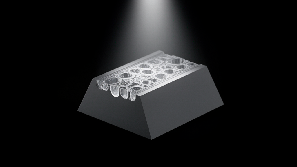
Cryogenic electron tomography of vitrified biological samples has emerged as an essential methodology for resolving cellular architecture in near-native environments at nanometer-scale resolution. To image structures inside intact cells, researchers employ cryogenic focused ion beam milling to produce thin, electron-transparent lamellae from frozen cellular material. However, conventional cryo-focused ion beam workflows rely on rectangular milling patterns that leave perpendicular retaining walls of unmilled cell remnants flanking the target area. Because a cryo-lamella possesses a flat slab geometry, sample tilting inside the transmission electron microscope is typically confined to an angular range of approximately plus or minus sixty degrees. This geometric constraint produces an angular gap in data collection known as the missing wedge in Fourier space. In reconstructed three-dimensional volumes, this missing wedge creates substantial resolution anisotropy, elongating structural features along the optical axis and obscuring fine details aligned perpendicular to the tilt axis.
To overcome this long-standing limitation, a research team led by Moritz Wachsmuth-Melm, Cornelis Mense, Santiago Gomez Melo, Liv Zimmermann, Ulrich S. Schwarz, and Petr Chlanda developed a trapezoidal milling geometry that unlocks dual-axis cryogenic electron tomography on cellular cryo-lamellae. Publishing their findings in the journal BMC Methods, the investigators engineered a micromachining approach using polygonal patterns within focused ion beam control software to eliminate obscuring cellular material. In standard rectangular milling geometries, the surrounding bulk cell body casts an electron-opaque shadow over the lamella at steep viewing angles, precluding data collection around a second, orthogonal tilt axis. The researchers calculated that the obscured region is geometrically governed by the tangent of the tilt angle multiplied by the surrounding cell height. By implementing two opposing isosceles trapezoid milling patterns whose shorter bases face one another, the team created open clearance geometries that prevent surrounding cell walls from clipping the electron beam during high-angle tilting.
The mathematical formulation of the trapezoid pattern required precise geometric dimensions to guarantee unobstructed transmission during dual-axis acquisition. For an isosceles trapezoid with shorter base b1, longer base b2, and height h, the longer base satisfies the relation where b2 equals b1 plus two times h multiplied by the cotangent of thirty degrees. To attain an accessible tilt angle of sixty degrees, the interior angle at the shorter base was established at one hundred and fifty degrees, complemented by a supplementary thirty-degree angle. In practical laboratory implementation on adherent Vero E6 cells plunge-frozen onto gold mesh grids, the investigators milled patterns with a shorter base of fourteen micrometers, a longer base of approximately thirty micrometers, and a milling height of 4.7 micrometers. Following coarse trapezoidal ablation, a secondary polygonal pattern comprising six vertices reduced unnecessary milling volume, after which sequential rectangular thinning and final polishing steps at grazing angles between eight and ten degrees brought lamellae to target thickness.
The experimental protocol was conducted using an Aquilos 2 cryogenic focused ion beam scanning electron microscope and a Titan Krios transmission electron microscope operated at three hundred kiloelectronvolts. Adherent Vero E6 cells were cultured on silicon dioxide coated gold grids, blotted for 3.5 seconds, and plunge-frozen in liquid ethane maintained at minus one hundred and eighty-five degrees Celsius. The resulting specimens were milled using a customized fifteen-hole aperture strip delivering beam currents systematically graded from five hundred to one thousand picoamperes down to thirty picoamperes for final polishing. To collect dual-axis tilt series, the team utilized a specialized stage inside the transmission electron microscope capable of ninety-degree in-plane specimen rotation. Cryo-TEM montaged maps confirmed that the trapezoid geometry cleared obstruction across approximately eighty percent of the lamella surface area during extreme specimen tilting, successfully resolving the shadowing that previously restricted cellular cryo-lamellae strictly to single-axis data acquisition schemes.
Data collection was performed using a high-resolution direct electron detector equipped with an energy filter operating at an energy slit width of fifteen electronvolts. Tilt series were recorded using parallel beam illumination and automated acquisition routines over an A-axis range spanning from plus sixty-eight degrees to minus fifty-two degrees and an orthogonal B-axis range spanning from plus sixty degrees to minus sixty degrees in three-degree increments. To prevent electron radiation damage from degrading high-resolution cellular structures across two full tilt series, the authors divided the cumulative electron dose between the two orthogonal directions. By comparing full-dose series with half-dose series generated through movie frame splitting, a two-sided paired t-test yielded a p-value of 0.166, demonstrating that reducing electron dose per tilt series produced no statistically significant increase in patch tracking alignment residual errors while maintaining robust fiducial alignment during tomographic reconstruction.
Reconstructed dual-axis tomograms were systematically compared against conventional single-axis volumes as well as single-axis tomograms processed with state-of-the-art computational missing wedge restoration algorithms, specifically IsoNet and DeepDeWedge. While deep learning algorithms aim to fill missing Fourier coefficients through trained neural network inference, direct qualitative and quantitative assessments revealed that computational restoration alone could not fully recover membrane boundaries and filamentous structures oriented perpendicular to the primary tilt axis. In contrast, physical dual-axis data acquisition directly recovered orthogonal spatial frequencies. Applying unsupervised automated three-dimensional membrane segmentations with the MemBrain software suite demonstrated superior continuity and clarity of complex intracellular membranes in dual-axis reconstructions. Furthermore, the authors noted that applying computational restoration to both axes prior to tomographic combination yielded enhanced structural fidelity, showing that physical dual-axis tilt series provide valuable experimental training data for developing future machine learning reconstruction architectures.
Beyond beam obstruction during imaging, cellular cryo-lamellae face severe mechanical vulnerabilities during microfabrication and sample handling. Cryo-lamellae must remain anchored to the surrounding cellular material, yet the release of internal compression during ion beam milling and strong thermal contraction mismatches frequently induce bending, wrinkling, or catastrophic mechanical fracture. To uncover the physical mechanisms governing lamella failure, the researchers constructed a linear elasticity simulation framework using the open-source software libraries Dolfinx and Gmsh. The vitrified cellular matrix was modeled as an isotropic elastic solid characterized by a Young’s modulus of eight gigapascals and a Poisson’s ratio of 0.15. Thermal strain was simulated across a temperature drop of two hundred Kelvin, evaluating differential contraction between the vitrified biological material, with a thermal expansion coefficient of two times ten to the negative fifth per Kelvin, and the underlying amorphous carbon support film.
Finite element simulations evaluated the spatial distribution of displacement fields and equivalent von Mises stresses across three distinct lamella junction designs: conventional orthogonal rectangular milling with sharp T-joints, ramp-step joints formed by stepwise narrowing of milling rectangles, and angled Y-joints produced by trapezoid milling. The numerical analyses revealed that sharp T-joints generate severe stress concentrations reaching values up to thirty megapascals, contrasted with baseline stresses between 0.1 and fifteen megapascals throughout the remaining cell volume. This stress accumulation at T-joints is driven by sharp reentrant contact angles combined with inward structural buckling under thermal contraction. Conversely, both ramp-step and Y-joint geometries suppressed local joint buckling by increasing separation between the lamella and adjacent cell walls, shifting von Mises stresses into the body of the lamella where values spanned from twenty to thirty-seven megapascals.
The computational simulations also identified critical limitations in conventional stress-relief designs when applied to newly developed lamella geometries. In standard rectangular milling, vertical stress-relief cuts—also called micro-expansion joints—effectively isolate the thin central foil by absorbing lateral contraction. However, the simulation results demonstrated that conventional vertical cuts provide diminished mechanical protection when paired with trapezoidal Y-joints because the geometric separation between the cuts and the lamella prevents stress dissipation. To mitigate this stress concentration, the team modeled alternative stress-relief geometries, including angled triangular cuts and V-shaped cuts positioned closer to the lamella boundary. Finite element calculations indicated that both triangular and V-shaped relief configurations significantly lowered von Mises stresses within the lamella foil. Translating these simulated designs to laboratory practice, the authors successfully milled three lamellae containing triangular cuts and three containing V-shaped cuts, with all six specimens surviving subsequent transfer to the transmission electron microscope intact.
The researchers further evaluated the mechanical influence of smoothing junction boundaries by varying the curvature radius of joints connecting the lamella to the bulk cell body. In simulations of rectangular geometries, introducing rounded fillets with low curvature radii dramatically reduced peak von Mises stresses at contact corners. Parametric modeling identified an optimal stress minimum at a curvature radius of approximately 0.32 micrometers. When the fillet radius was expanded further up to 1.45 micrometers, the additional vitrified material increased overall stiffness and reversed the protective effect, elevating stresses across the lamella back toward twenty megapascals. Similarly, simulating an asymmetric design featuring a T-joint on one edge and a cut-through notch on the opposing edge—an approach derived from waffle milling protocols—demonstrated effective strain relief by eliminating rigid bilateral mechanical constraint.
Together, these experimental and computational findings establish a comprehensive framework for optimizing both information content and mechanical throughput in cellular cryo-electron tomography workflows. The trapezoid milling strategy permits direct dual-axis data acquisition across eighty percent of lamella surfaces without cell body shadowing, providing experimental ground truth that resolves missing wedge distortions and improves automated segmentation of crowded organelles. Simultaneously, finite element analysis elucidates how thermal contraction mismatches between vitreous ice and supporting substrates generate structural fractures, guiding the rational placement of V-shaped relief cuts and rounded connection fillets. The authors conclude that integrating automated polygonal focused ion beam milling with mechanical stress modeling will enhance sample survival rates and accelerate the adoption of dual-axis tomography in structural cell biology.
Subject of Research: Cryo-electron tomography methods and mechanical modeling of cellular cryo-lamellae
Article Title: On-lamella dual-axis cryo-electron tomography and modelling of lamella stability
Article References: Wachsmuth-Melm, M., Mense, C., Gomez Melo, S., Zimmermann, L., Schwarz, U. S., & Chlanda, P. (2026). On-lamella dual-axis cryo-electron tomography and modelling of lamella stability. BMC Methods, 3(1), Article 5. https://doi.org/10.1186/s44330-026-00056-9
Image Credits: AI Generated
DOI: 10.1186/s44330-026-00056-9
Keywords: cryo-electron tomography, cryo-FIB milling, structural biology, finite element method, cellular imaging, dual-axis tomography, von Mises stress, electron microscopy, On-lamella, dual-axis, cryo-electron, tomography
Cite Scienmag News
Gregory Coleman. (November 9, 2026). Trapezoid Cryo-FIB Milling Enables Dual-Axis Cellular Tomography and Lamella Stability. Scienmag. https://scienmag.com/trapezoid-cryo-fib-milling-enables-dual-axis-cellular-tomography-and-lamella-stability/
Gregory Coleman. "Trapezoid Cryo-FIB Milling Enables Dual-Axis Cellular Tomography and Lamella Stability." Scienmag, 9 November 2026, https://scienmag.com/trapezoid-cryo-fib-milling-enables-dual-axis-cellular-tomography-and-lamella-stability/. Accessed 9 November 2026.
Gregory Coleman. "Trapezoid Cryo-FIB Milling Enables Dual-Axis Cellular Tomography and Lamella Stability." Scienmag. November 9, 2026. https://scienmag.com/trapezoid-cryo-fib-milling-enables-dual-axis-cellular-tomography-and-lamella-stability/




