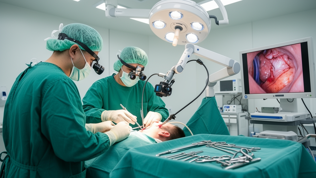
Trigeminal neuralgia, a severe craniofacial pain disorder most often caused by neurovascular compression, is usually treated surgically with microvascular decompression. A new systematic review and meta-analysis published in Acta Neurochirurgica compares two ways of performing that operation: the traditional microscopic approach and a newer endoscopic one. The analysis, which followed PRISMA guidelines and searched PubMed, Embase, and Web of Science for comparative studies, pooled data from ten studies covering 1,051 patients, of whom 465 underwent endoscopic microvascular decompression and 586 the microscopic procedure.
The surgical rationale for the comparison rests on optics. Microscopic microvascular decompression remains the gold-standard surgical treatment for the condition, but its linear field of view may limit visualization of the offending vessels pressing on the trigeminal nerve. The endoscopic technique offers enhanced illumination and panoramic visualization, which could in principle allow more complete decompression and fewer complications. Because comparative data across studies had remained inconsistent, the authors set out to compare the efficacy and safety of the two approaches directly.
On the primary question of pain control, the pooled results showed little to separate the operations. Adequate pain relief was comparable between the two approaches at all measured time points. Where the endoscopic approach did appear to pull ahead was in the strictest measure of success: complete long-term pain freedom favored endoscopic surgery, with an odds ratio of 2.72 and a p-value below 0.01.
Recurrence also differed between the groups. Patients treated endoscopically had significantly lower recurrence of pain, with an odds ratio of 0.51 and a p-value of 0.02. Overall complications were reduced with the endoscopic approach as well, at an odds ratio of 0.48 with p below 0.001. This overall difference was driven particularly by postoperative headache, which was markedly less frequent after endoscopic surgery, with an odds ratio of 0.28 and p below 0.001; other individual complications were comparable between the two techniques.
The analysis also captured differences inside the operating room. Endoscopic procedures more frequently detected multiple offending vessels, with an odds ratio of 1.99 and p equal to 0.035, a finding consistent with the idea that panoramic visualization reveals vascular compressions that a linear microscopic view can miss. Operative time showed a marginal difference of 6.4 minutes favoring the microscopic approach, while decompression time differed marginally by 2.8 minutes in the other direction, both with p-values below 0.05. Pooled estimates across all outcomes were calculated using a random-effects model, with heterogeneity assessed using the I2 statistic.
The authors are careful about how far these numbers can be taken. The included studies were non-randomized, and the certainty of the evidence was low. The review therefore concludes that endoscopic microvascular decompression appears to provide pain relief comparable to the microscopic operation, with studies suggesting possible advantages in long-term complete pain relief, recurrence, and postoperative headache, but that these associations cannot establish superiority or a causal effect of the endoscopic approach.
Instead, the findings are framed as hypothesis-generating. The review calls for confirmation in prospective, standardized studies, ideally randomized or well-adjusted multicenter trials with long-term follow-up, before any claim of superiority can be made.
For surgeons and patients weighing options, the practical takeaway is nuanced. The endoscopic technique did not shorten operations meaningfully and did not reduce complications across the board, but it was associated with better detection of multiple compressing vessels, less postoperative headache, less recurrence, and more complete long-term pain freedom in the pooled observational data. Whether those associations reflect genuine advantages of the endoscope’s illumination and panoramic view, or differences in patient selection, surgeon experience, or study design across the ten included studies, is precisely the question that randomized trials would need to answer.
Subject of Research: A systematic review and meta-analysis comparing endoscopic and microscopic microvascular decompression for trigeminal neuralgia.
Article Title: Endoscopic versus microscopic microvascular decompression for trigeminal neuralgia: a systematic review and meta-analysis
Article References: Punukollu, A., Mendieta-Barrera, C. D., Lobo, K., Koppanatham, A., Santos, J. G. C. S. M., Hwang, H. S., e Silva, P. M. P. B. R., Schevz, R., Ghaffari-Rafi, A., Tripathi, M., & Prevedello, D. (2026). Endoscopic versus microscopic microvascular decompression for trigeminal neuralgia: a systematic review and meta-analysis. Acta Neurochirurgica. https://doi.org/10.1007/s00701-026-07045-5
Image Credits: AI Generated
DOI: 10.1007/s00701-026-07045-5
Keywords: trigeminal neuralgia, microvascular decompression, endoscopic surgery, meta-analysis, neurosurgery, neurovascular compression, postoperative headache, pain relief, random-effects model, systematic review, Endoscopic, versus
Cite Scienmag News
Ophelia Keating. (October 29, 2026). Endoscopic Microvascular Decompression Shows Possible Edge Over Microscopic Surgery. Scienmag. https://scienmag.com/endoscopic-microvascular-decompression-shows-possible-edge-over-microscopic-surgery/
Ophelia Keating. "Endoscopic Microvascular Decompression Shows Possible Edge Over Microscopic Surgery." Scienmag, 29 October 2026, https://scienmag.com/endoscopic-microvascular-decompression-shows-possible-edge-over-microscopic-surgery/. Accessed 29 October 2026.
Ophelia Keating. "Endoscopic Microvascular Decompression Shows Possible Edge Over Microscopic Surgery." Scienmag. October 29, 2026. https://scienmag.com/endoscopic-microvascular-decompression-shows-possible-edge-over-microscopic-surgery/